Apoptosis-Based Evaluation Of Chemosensitivity In Cancer Patients

a cancer patient and chemosensitivity technology, applied in the field of cancer therapies and prediction of tumor cell response to chemotherapeutic agents, can solve the problems of inability to meet clinical situations, no reliable means of early detection available,

Active Publication Date: 2007-11-29
YALE UNIV
View PDF0 Cites 3 Cited by
  • Summary
  • Abstract
  • Description
  • Claims
  • Application Information

AI Technical Summary

Problems solved by technology

Nearly 75% of patients present with advanced disease since, as of yet, no reliable means for early detection is available.
Such assays are reasonably predictive of drug action in vivo and remain the standard against which other assays are compared but the best of them are not amenable to clinical situations given the low clonogenic efficiencies of most primary tumor samples in culture.

Method used


Image

  • Apoptosis-Based Evaluation Of Chemosensitivity In Cancer Patients
  • Apoptosis-Based Evaluation Of Chemosensitivity In Cancer Patients
  • Apoptosis-Based Evaluation Of Chemosensitivity In Cancer Patients

Examples


examples

Methods

Cell Culture

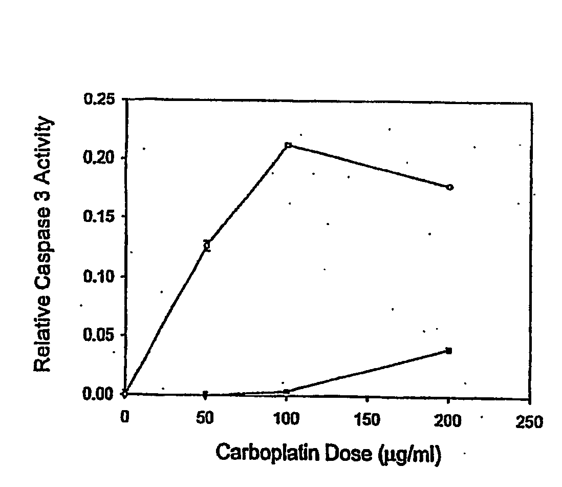
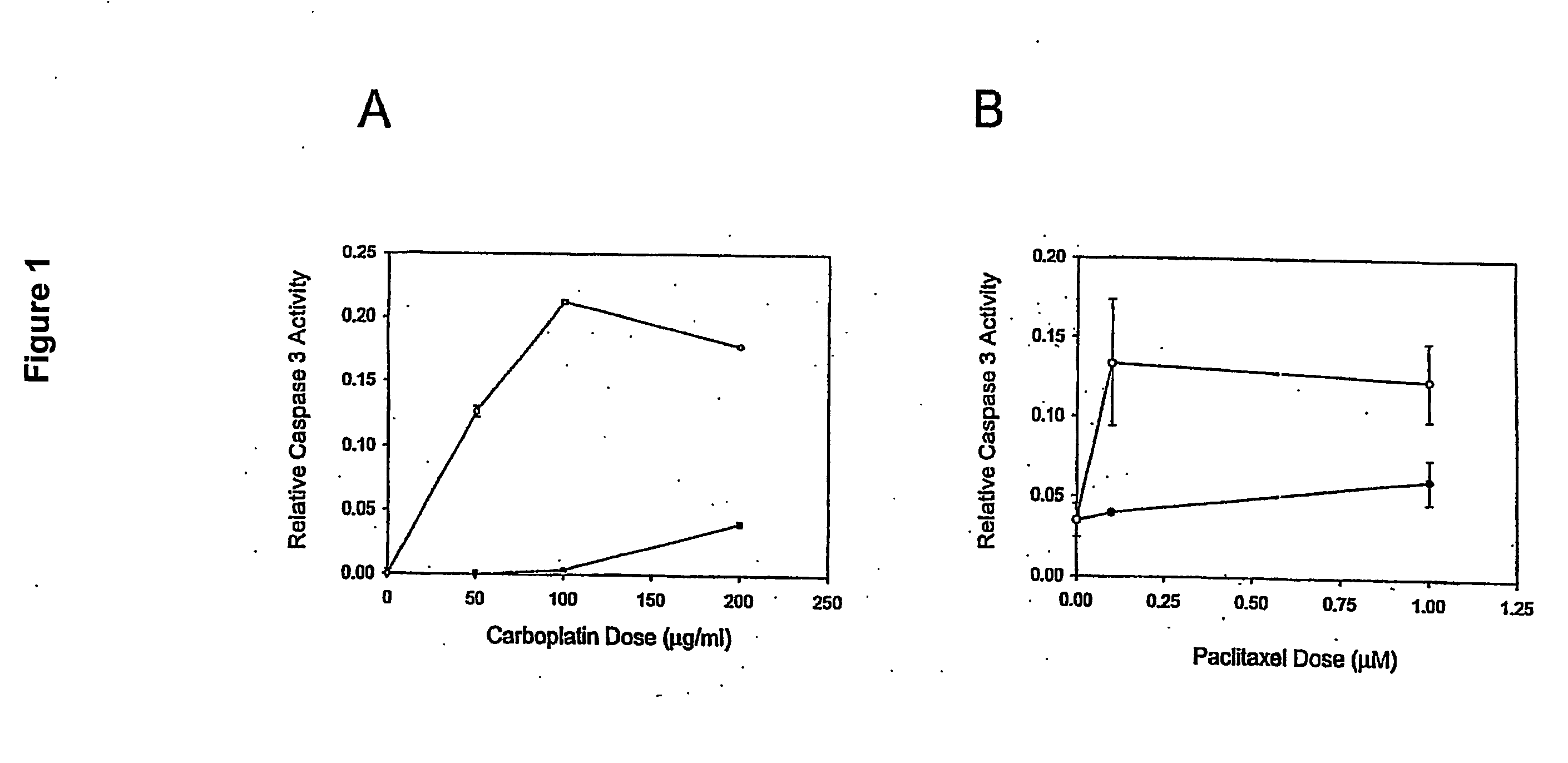
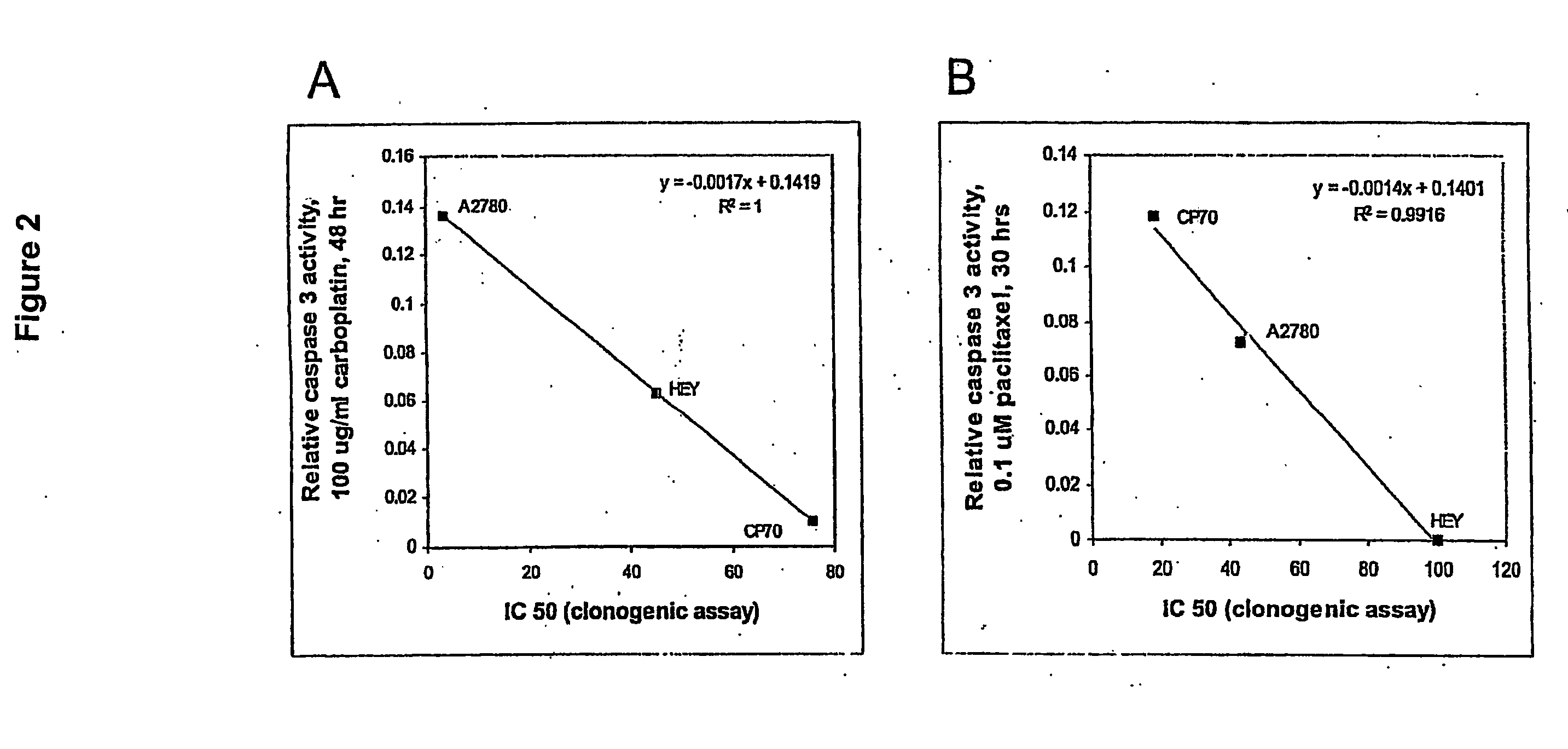
[0020] Ovarian cancer cell lines HEY (gift from Dr. J M Trent, (26)), A2780 and CP70 (gifts from Dr. T C Hamilton, (27)) were cultured at 37° C. in a 5% CO2 atmosphere. Culture medium varied with the cell line: DME / F12 plus 1% calf serum (HEY); DME / F12 plus 10% FBS, 584 ug / ml L glutamine (A2780 and CP70). All formulations contained 100 μg / ml streptomycin, 62.5 μg / ml penicillin and 2.5 μg / ml amphotericin. Primary cells were cultured in 50% 199 media and 50% MCDB 105 media (Sigma), supplemented with 10% fetal bovine serum (Hyclone, South Logan, Utah), 10 mM HEPES, 0.1 mM MEM non-essential amino acids, 1 mM sodium pyruvate, 100 nM penicili / streptomycin (Gibco) and 4 ng / ml EGF (Sigma). For drug treatments, approximately 1×10⁵ cells in exponential growth were sub-cultured in a 25 cm2 flask per treatment. Carboplatin (Sigma C2538) or paclitaxel (Sigma T7402) was added to the media after the cells reached 60-70% confluence as described in the Results.

Patient Samples...


PUM

PropertyMeasurementUnit
length of timeaaaaaaaaaa
pHaaaaaaaaaa
pHaaaaaaaaaa

Abstract

Induction of apoptosis in target cells is a key mechanism by which chemotherapy induces cell killing. An in vitro system has been established for determining carboplatin and paclitaxel (Taxol) chemosensitivity of epithelial ovarian cancer cells, where measurements of caspase-3 activation are surrogate markers for activation of chemotherapy-induced programmed cell death. To validate the assay as a predictor of clinical chemotherapy-induced programmed cell death. To validate the assay as a predictor of clinical chemosensitivity in vitro apoptotic response were compared to the clinical response of the patients from whom the tumor cells were isolated. Caspase-3 activation in response to in vitro chemotherapy to both drugs was shown to have an 83% positive predictive value and a 71% negative predictive value. Markers of apoptosis such as caspase-3 activation can be quantitated and utilized to predict the clinical response to chemotherapy.

Description

CROSS-REFERENCE TO RELATED APPLICATIONS [0001] This application claims priority to U.S. provisional patent application no. 60 / 525,220, filed Nov. 26, 2003, incorporated by reference herein in its entirety.STATEMENT REGARDING FEDERALLY-SPONSORED RESEARCH OR DEVELOPMENT [0002] This work was supported by grants from the Ethyl F. Donaghue Foundation and by grants from the National Institutes of Health, RO1 HD37137-01A2 and R01CA92435-01.FIELD OF THE INVENTION [0003] The invention is in the field of cancer therapies and prediction of tumor cell response to chemotherapeutic agents. BACKGROUND OF THE INVENTION [0004] Over 14,000 women in the U.S., each year die from epithelial ovarian carcinomas (1). Nearly 75% of patients present with advanced disease since, as of yet, no reliable means for early detection is available. The effective treatment of advanced or recurrent ovarian cancer is often compromised by both the intrinsic and acquired resistance of many ovarian carcinomas to one or mor...

Claims


Application Information

Patent Timeline
Patent Type & Authority Applications(United States)
IPC IPC(8): C12Q1/02G01N1/30G01N33/48G01N33/50G01N33/574
CPCG01N1/30G01N2510/00G01N2333/96466G01N33/5011
Inventor MOR, GUILLEMO
Owner YALE UNIV
Why Eureka
Try Eureka