Systems and Methods for Automated Segmentation, Visualization and Analysis of Medical Images

a technology of medical images and automated segmentation, applied in the field of systems and methods for aiding in medical diagnosis and evaluation of internal organs, can solve the problems of difficult diagnosis of stenosis or other abnormalities, and inability to provide efficient or intuitive means to navigate through virtual organs

Inactive Publication Date: 2007-11-29
VIATRONIX
View PDF5 Cites 207 Cited by
  • Summary
  • Abstract
  • Description
  • Claims
  • Application Information

AI Technical Summary

Benefits of technology

[0005] The present invention is directed to systems and methods for visualization and navigation of complex 2D or 3D data models of internal organs, and other components, which models are generated from 2D image datasets produced by a medical imaging acquisition device (e.g., CT, MRI, etc.). In one exemplary embodiment, an imaging system for automated segmentation and visualization of medical images includes an image processing module for automatically processing image data using a set of directives to identify a target object in the image data and process the image data according to a specified protocol, a rendering module for automatically generating one or more images of the target object based on one or more of the directives and a digital archive for storing the one or more generated images. The image data may be DICOM-formatted image data, wherein the imaging processing module extracts and processes meta-data in DICOM fields of the image data to identify the target object. The image processing module directs a segmentation module to segment the target object using processing parameters specified by one or more of the directives.

Problems solved by technology

However, while stacks of such slices may be useful for analysis, they do not provide an efficient or intuitive means to navigate through a virtual organ, especially one as tortuous and complex as the colon, or arteries.
For example, when imaging blood vessels, cross-sections merely show slices through vessels, making it difficult to diagnose stenosis or other abnormalities.

Method used


Image

  • Systems and Methods for Automated Segmentation, Visualization and Analysis of Medical Images
  • Systems and Methods for Automated Segmentation, Visualization and Analysis of Medical Images
  • Systems and Methods for Automated Segmentation, Visualization and Analysis of Medical Images

Examples


Embodiment Construction

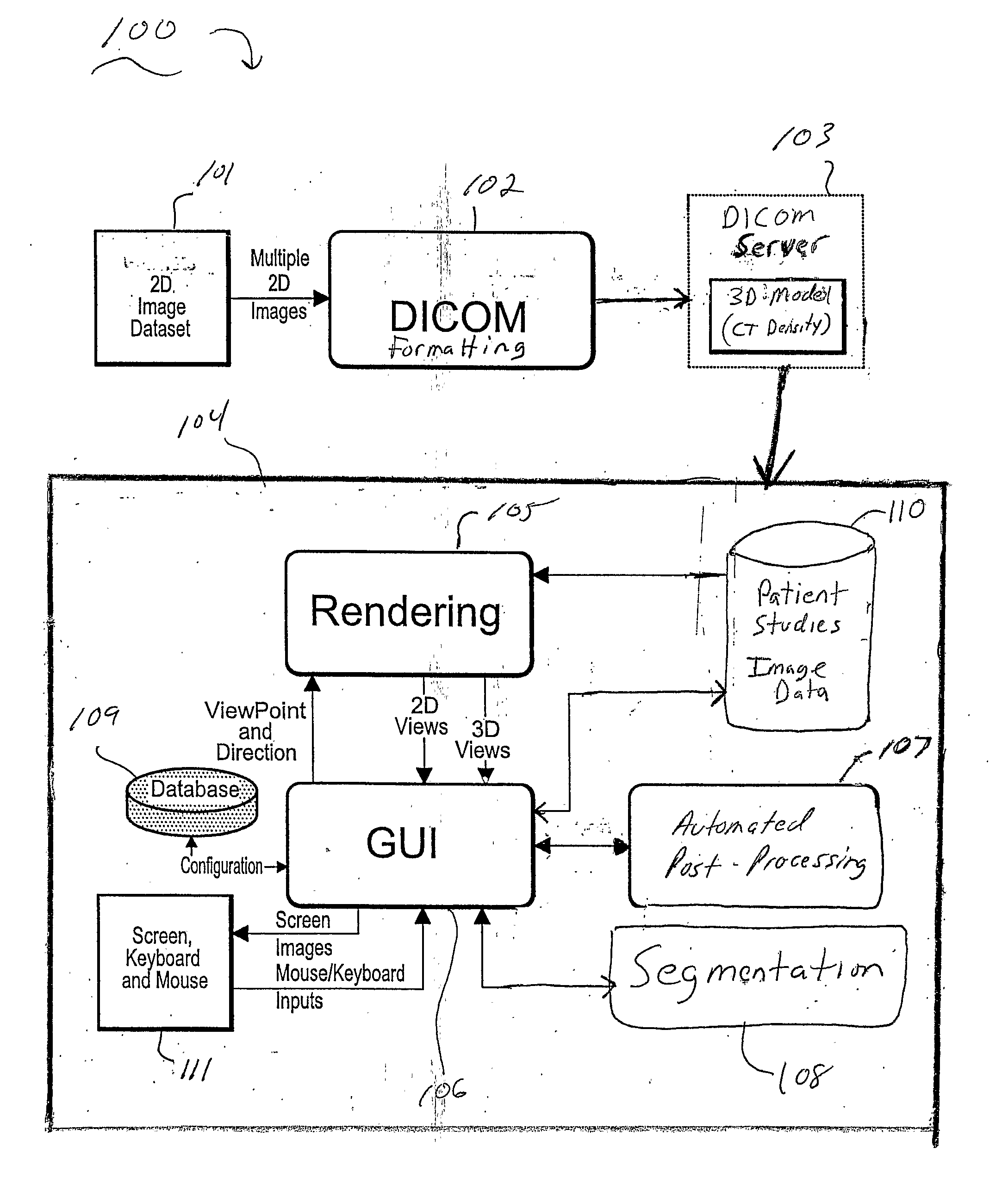
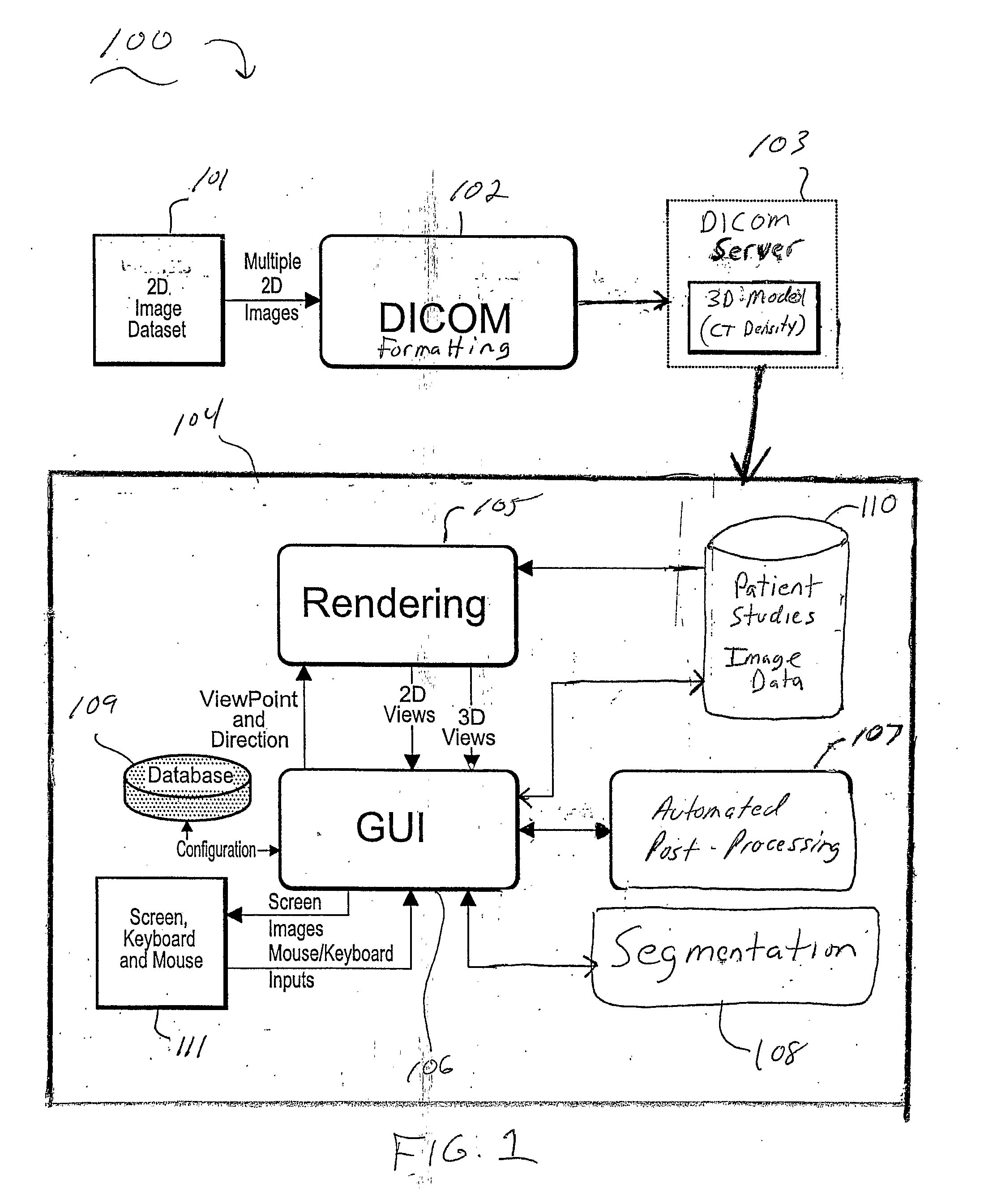
[0012] The present invention is directed to medical imaging systems and methods for assisting in medical diagnosis and evaluation of a patient. Imaging systems and methods according to preferred embodiments of the invention enable visualization and navigation of complex 2D and 3D models of internal organs, and other components, which are generated from 2D image datasets generated by a medical imaging acquisition device (e.g., MRI, CT, etc.).

[0013] It is to be understood that the systems and methods described herein in accordance with the present invention may be implemented in various forms of hardware, software, firmware, special purpose processors, or a combination thereof. Preferably, the present invention is implemented in software as an application comprising program instructions that are tangibly embodied on one or more program storage devices (e.g., magnetic floppy disk, RAM, CD ROM, DVD ROM, ROM and flash memory), and executable by any device or machine comprising suitable ...


PUM


Abstract

An imaging system for automated segmentation and visualization of medical images (100) includes an image processing module (107) for automatically processing image data using a set of directives (109) to identify a target object in the image data and process the image data according to a specified protocol, a rendering module (105) for automatically generating one or more images of the target object based on one or more of the directives (109) and a digital archive (110) for storing the one or more generated images. The image data may be DICOM-formatted image data (103), wherein the imaging processing module (107) extracts and processes meta-data in DICOM fields of the image data to identify the target object. The image processing module (107) directs a segmentation module (108) to segment the target object using processing parameters specified by one or more of the directives (109).

Description

CROSS-REFERENCE TO RELATED APPLICATIONS [0001] This application claims priority to U.S. Provisional Application No. 60 / 525,603, filed Nov. 26, 2003, and U.S. Provisional Application No. 60 / 617,559, filed on Oct. 9, 2004, which are fully incorporated herein by reference.TECHNICAL FIELD OF THE INVENTION [0002] The present invention relates generally to systems and methods for aiding in medical diagnosis and evaluation of internal organs (e.g., blood vessels, colon, heart, etc.) More specifically, the invention relates to a 3D visualization system and method for assisting in medical diagnosis and evaluation of internal organs by enabling visualization and navigation of complex 2D or 3D data models of internal organs, and other components, which models are generated from 2D image datasets produced by a medical imaging acquisition device (e.g., CT, MRI, etc.). BACKGROUND [0003] Various systems and methods have been developed to enable two-dimensional (“2D”) visualization of human organs ...

Claims


Application Information

Patent Timeline
Patent Type & Authority Applications(United States)
IPC IPC(8): A61B5/00A61B5/05A61B8/00G06FG06T7/00G06T17/40
CPCG06T7/0012G06T2207/30004G06T2219/008G06F19/321G06T19/00G06T2210/41G16H30/20G16H30/40
Inventor DACHILLE, FRANK C.CHEN, DONGQINGMEISSNER, MICHAELCAI, WENLI
Owner VIATRONIX
Why Eureka
Try Eureka