Apparatus and method for spectrally measuring fundus

a technology of spectroscopic fundus and measuring apparatus, which is applied in the field of spectroscopic fundus measuring apparatus, can solve the problems of difficult to perform accurate analyses, unable to achieve hyper-spectral light separation without burdening humans, and most studies carried out to date are far from spectral image measurement in full-scale, so as to avoid the choice of improper images and achieve clearest images. the effect of choosing efficiently

Inactive Publication Date: 2008-01-10
KK TOPCON
View PDF2 Cites 24 Cited by
  • Summary
  • Abstract
  • Description
  • Claims
  • Application Information

AI Technical Summary

Benefits of technology

[0012] Here, a series of spectral fundus images of different wavelengths typically mean a group of spectral fundus images of the same subject eye photographed in succession while increasing or decreasing the wavelength in a predetermined wavelength range. Changes in the photographing order or slight changes in the photographing conditions are acceptable. Processing means image processing including position matching correction among spectral fundus images, transformation such as projective transformation, filtering such as noise removal and edge detection, and expanding and thinning a line. The predetermined wavelength ranges typically mean ranges in which relatively clear images of respective specific parts can be obtained. The ranges may be determined by measuring the contrasts between the specific parts and their backgrounds or according to an empirical rule. The specific parts mean distinctive parts in a fundus image such as retinal arteries, retinal veins, optic nerve head, choroid, and macula area. With the above constitution, there can be provided a spectroscopic fundus measuring apparatus capable of identifying each part in spectral fundus images easily and accurately based on its spectral characteristic.
[0014] With the above constitution, a group of clear images can be preliminarily extracted for each specific part and the clearest image is chosen therefrom. Thus, the clearest image can be chosen efficiently and the choice of an improper image can be avoided.
[0028] With this constitution, the retinal arteries and the retinal veins can be distinguished efficiently.
[0031] The invention related to aspect (12) is the spectroscopic fundus measuring method related to aspect (11), as shown in FIG. 4 for example, wherein the image processing step has an image choosing step (S007) of choosing one of the extracted spectral fundus images or one of images of each of divided areas in the extracted spectral fundus images for each of the specific part as the clearest image, and, in the case where one of images of each of divided areas is chosen as the clearest image in the image choosing step, an image connecting step (S008) of connecting the clearest images of all the areas to form the clearest images of the entire fundus. With the above constitution, a group of clear images can be preliminarily extracted for each specific part and the clearest image is chosen therefrom. Thus, the clearest image can be chosen efficiently and the choice of an improper image can be avoided.
[0032] According to the present invention, there can be provided a spectroscopic fundus measuring apparatus capable of identifying each part in spectral fundus images based on its spectral characteristic easily and accurately and a measuring method therefor.

Problems solved by technology

However, most of the studies carried out up to now are far from spectral image measurement in full-scale.
While the hyper-spectral imaging is a technique in the spotlight and is used to obtain spectral images of the fundus, it is hard to perform accurate analyses because the amount of light of spectral images obtained varies greatly by the wavelength.
Moreover, the hyper-spectral light separation with a light amount without putting burden on humans has yet to be realized.
However, as restricted by for example the wavelength tunable time of the liquid crystal wavelength tunable filter and the exposure time of the camera, it takes about 20 seconds to take images at every 10 nm in the wavelength range from 500 nm to 720 nm.
Because alignment between the eye and the apparatus varies during that time, there has been another problem that the spectral images taken of the same part are displaced from each other.

Method used


Image

  • Apparatus and method for spectrally measuring fundus
  • Apparatus and method for spectrally measuring fundus
  • Apparatus and method for spectrally measuring fundus

Examples


first embodiment

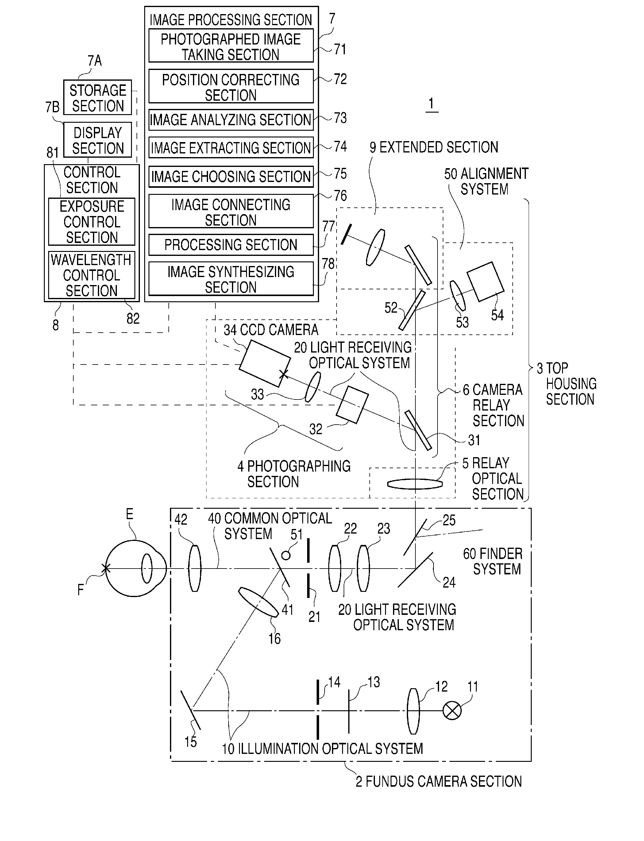
[0050]FIG. 1 shows a general example of an optical system of a spectral fundus image data measuring apparatus 1 as an embodiment of the invention. In the drawing, the spectral fundus image data measuring apparatus 1 may be roughly divided into: a fundus camera section 2, a top housing section 3, an image processing section 7, a storage section 7A, a display section 7B, and a control section 8. The fundus camera section 2 comprises: an illumination optical system 10 for illuminating the fundus F of a subject eye E, the fore stage section of a light receiving optical system 20 for receiving light beam reflected from the fundus F and forming a fundus image on the light receiving surface of a photographing section 4, a finder optical system 60 for an optometrist to observe the fundus F, etc. The top housing section 3 is made up of: the photographing section 4 for photographing a spectral fundus image, an alignment optical system 50 for aligning the illumination position of the illuminat...

second embodiment

[0082] An example in which each fundus image is divided into a plurality of areas, the clearest image with the highest contrast is chosen from images of each area in a plurality of extracted images, and the chosen images are connected to form the clearest images of the entire fundus is described in the first embodiment. In the second embodiment, an example in which the clearest image is manually chosen from images of each area in the extracted images is described. A plurality of extracted images are displayed on the display section 7B. When the operator designates a part in each of the areas, the spectral characteristic and the contrast of the part are displayed. The operator compares the spectral characteristic with the standard spectral characteristic of the specific part to confirm that the part belongs to the specific part, and chooses an extracted image with the highest contrast in a plurality of extracted images as the clearest image for the area. The clearest images chosen fo...

third embodiment

[0083] An example in which the clearest image is manually chosen from the images of each area is described in the second embodiment. In the third embodiment, an example in which a plurality of extracted images are manually compared with one another to choose the clearest image of the entire fundus is described. Also in this case, when the operator designates a part in an extracted image, the spectral characteristic and contrast of the part are displayed. The operator compares the spectral characteristic with the standard spectral characteristic of a specific part to confirm that the part belongs to the specific part, and chooses an extracted image with the highest contrast from a plurality of extracted images as the clearest image. For example, an image of retinal arteries and veins is chosen from images of a wavelength of 570 nm, an image of optic nerve head from images of a wavelength of 640 nm, and an image of choroidal vessels from images of a wavelength of 700 nm as the cleares...


PUM


Abstract

To provide a spectroscopic fundus measuring apparatus capable of identifying each part in spectral fundus images easily and accurately based on its spectral characteristic and a measuring method therefor. A spectral fundus image measuring apparatus 1 of the present invention includes: an illumination optical system 10 having an illumination light source 11 for illuminating a fundus; a light receiving optical system 20 for receiving a wavelength-tunable light beam reflected from the illuminated fundus to photograph a series of spectral fundus images of different wavelengths; an image processing section 7 for processing the spectral fundus images; a storage section 7A for storing the spectral fundus images; and a display section 7B for displaying the spectral fundus images. The image processing section 7 has a position correcting section 72 for correcting the series of spectral fundus images photographed by the light receiving optical system 20 to match the positions of the same parts therein, and an image extracting section 74 for extracting spectral fundus images in wavelength ranges predetermined for respective specific parts from the series of spectral fundus images corrected in the position correcting section 72.

Description

BACKGROUND OF THE INVENTION [0001] 1. Technical Field [0002] The present invention relates to a spectroscopic fundus measuring apparatus and a measuring method therefor. In particular, the present invention relates to a spectroscopic fundus measuring apparatus that acquires spectral fundus images to facilitate identification of parts with different spectral characteristics and that can form an image in which every part is clear in consideration of the spectral characteristics thereof, and a measuring method therefor. [0003] 2. Related Art [0004] Fundus observation is doubtless important in ophthalmic diagnosis. At present, anomaly findings are obtained by diagnosing the eye fundus by means of colored fundus images, fluorescent contrast images, etc. from a fundus camera. If it is possible to measure quantitatively oxygen saturation degree on the fundus and constituent substances distributed in the retina, there is a possibility of finding out the functions of fine parts of the retina...

Claims


Application Information

Patent Timeline
Patent Type & Authority Applications(United States)
IPC IPC(8): A61B3/14
CPCA61B3/10
Inventor MIHASHI, TOSHIFUMIHIROHARA, YOKOAOKI, HIROYUKITSURUGA, YASUKO
Owner KK TOPCON
Why Eureka
Try Eureka