Spinal diagnostic methods and apparatus

a diagnostic method and apparatus technology, applied in the field of spinal diagnostic methods, can solve problems such as back pain of patients, and achieve the effect of alleviating pain, facilitating diagnosis and in some cases treatment of discogenic pain

Active Publication Date: 2008-01-10
GLOBUS MEDICAL INC
View PDF25 Cites 71 Cited by
  • Summary
  • Abstract
  • Description
  • Claims
  • Application Information

AI Technical Summary

Benefits of technology

[0013] Methods and devices of the present invention generally facilitate diagnosis, and in some cases treatment, of discogenic pain. More specifically, methods and devices of the invention help determine whether one or more intervertebral discs in a patient are actually causing the patient's back pain, and also help pinpoint which disc or discs are causing the pain. In one embodiment, a distal portion of a catheter device is positioned transannularly or transosseously in an intervertebral disc that is thought to be the cause of the patient's pain. One or more anchoring members are then used to maintain the distal portion in the disc. The patient is asked to assume a position or perform a task, such as bending over, which typically causes the patient to experience back pain. Using the catheter device, one or more substances, such as an anesthetic or analgesic, are injected into the disc. The patient then reports whether the anesthetic has alleviated the pain. Optionally, additional intervertebral discs may be tested, one or more placebo injections may be used, a conventional discography may be added to the procedure and / or the like. Based on the patient's response to the introduction of substance(s) into the disc (or discs), a determination may be made as to whether one or more specific discs are causing the patient's pain. Diagnostic and treatment decisions may be based on such a determination.
[0017] In various embodiments in which an introducer device is used, the introducer device may either be advanced to a position within the disc or to a position just outside the disc. In either case, the catheter device may have a distal end or portion configured to facilitate advancement of the distal end through the annulus fibrosis. In some embodiments, the catheter device is passed over a guidewire. In some cases, the catheter is passed over the guidewire within the needle, while in alternative embodiments the introducer is removed over the guidewire before the catheter device is passed over the guidewire. In some embodiments, positioning the distal portion is facilitated by visualizing at least one radioopaque marker or material at or near the distal portion to assess its location.
[0020] In addition to the anchor mechanisms described above, the access and treatment catheters of the present invention can be modified to promote tissue growth on or into at least selected portions of the catheter. Such tissue growth promotion could be accomplished by modifying the exterior texture or shape of the catheter exterior, or by providing additional physical structure which would promote tissue ingrowth and attachment. Alternatively or in addition, the catheter could be provided with an osteogenic substance, drug, or chemical to promote the desired tissue attachment. Such tissue attachment can be promoted in bone, muscle, fibrotic tissue, scar tissue, chondrocytes, or other tissue which occurs in or around the disc space of vertebral bodies. Anchoring by promoting tissue ingrowth is particularly useful when the catheter will be implanted for an extended length of time, as will often be the case for therapeutic treatment protocols.
[0030] A number of features may facilitate passage of the catheter body through an introducer device. For example, in one embodiment the catheter body includes a friction resistant outer surface. In some embodiments, the outer diameter of the catheter body is less than 2 mm. Also in some embodiments, a cross-sectional diameter of the catheter body is larger near a proximal end of the body than near a distal end of the body. Optionally, the catheter body may also include an outer surface having one or more markings for indicating depth of insertion of the catheter device into a patient's body. In alternative embodiments, the catheter body may include an outer surface having two or more different colors for indicating depth of insertion of the catheter device into a patient's body. The catheter body may further include at least one radioopaque marker or material for facilitating visualization of the catheter device in a patient.
[0033] It will also be preferred in some instances to provide a guidewire with radioopaque markers. The guidewire will have a small diameter, and superelastic materials, such as nitinol, have low visibility under fluoroscopic imaging. The radioopacity of the guidewire can be improved by adding a radiopaque material in a variety of conventional ways. For example, the guidewire can be coated with the radioopaque material, a radioopaque core may be provided within the guidewire, a radioopaque material may be braided over the guidewire, or the material provided as ring-like markers. Such coated guidewires having improved radioopacity can be manufactured in a variety of ways, including sputtering, electroplating, electroless plating, and the like.
[0039] In various embodiments, the kit may also include one or more additional devices, such as but not limited to an introducer device for facilitating positioning of the catheter device in the disc, a pointed stylet removably disposed within a lumen of the catheter device for piercing through the annulus fibrosis and / or a guidewire passable through the needle.

Problems solved by technology

The patient is asked to assume a position or perform a task, such as bending over, which typically causes the patient to experience back pain.

Method used


Image

  • Spinal diagnostic methods and apparatus
  • Spinal diagnostic methods and apparatus
  • Spinal diagnostic methods and apparatus

Examples


Embodiment Construction

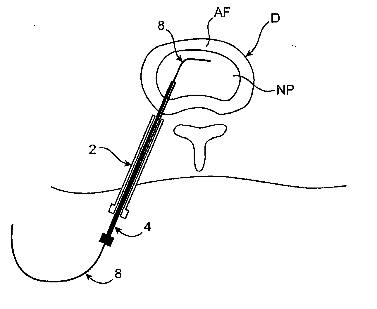
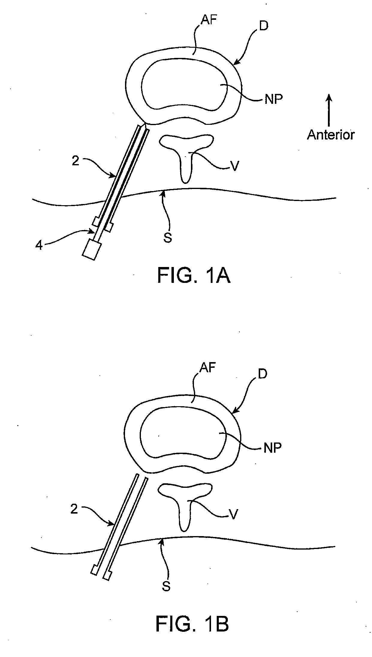
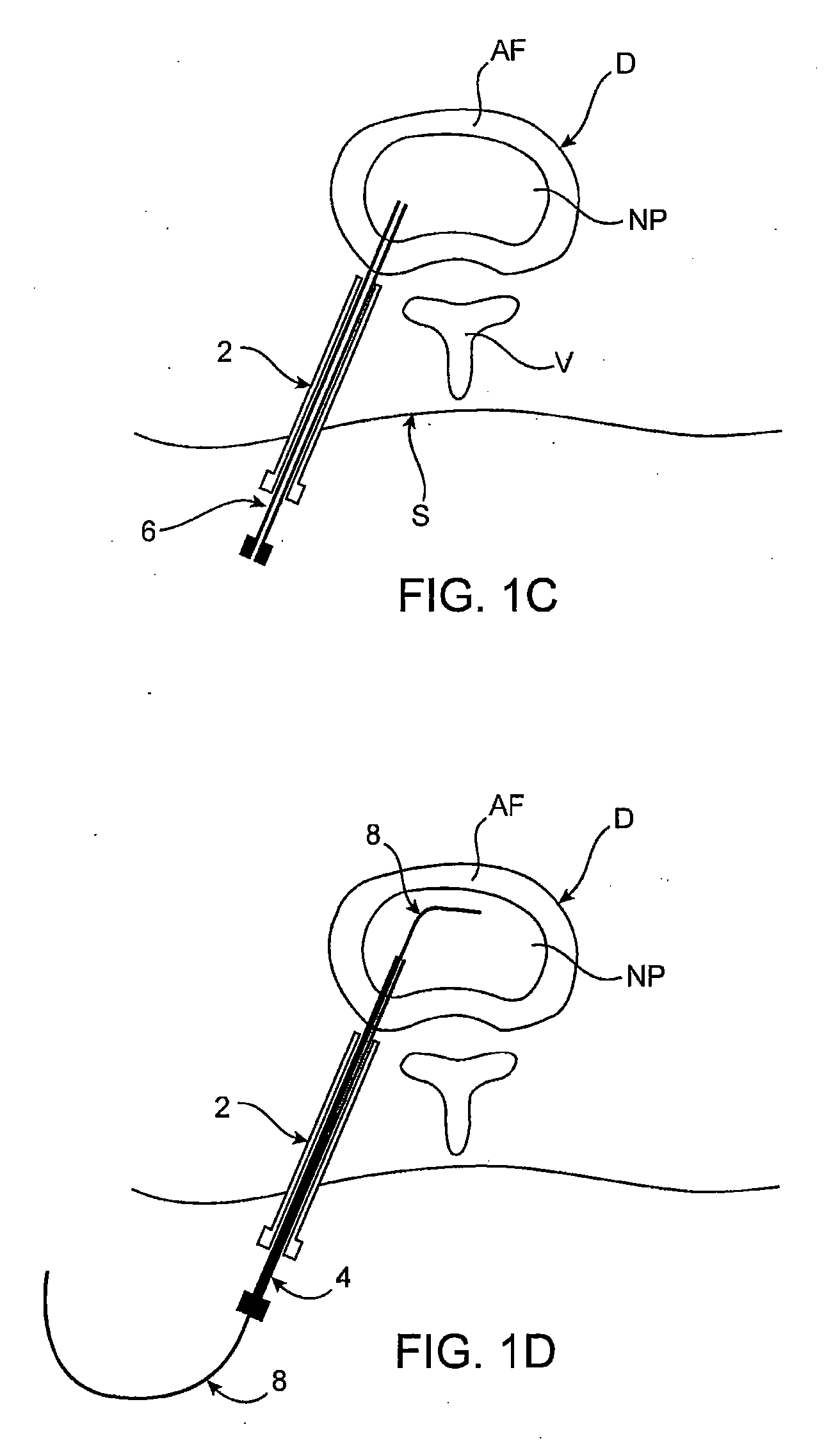
[0067] Methods, devices and systems of the present invention generally provide for introduction of one or more substances into an intervertebral disc to facilitate diagnosis and / or treatment of discogenic pain (i.e., back pain originating in one or more intervertebral discs). Methods, devices and systems may be used alone or in conjunction with other methods or devices that are currently known or hereafter developed, such as discography, radiological studies, physical examination and / or the like. In alternative embodiments, methods and devices of the invention may be used for purposes other than diagnosis or treatment, such as for study or experimental purposes or the like. Therefore, although the following description focuses on diagnostic and therapeutic applications, various embodiments may be used for any other suitable application.

[0068] Referring now to FIGS. 1A-1K, a method for introducing a substance into an intervertebral disc is illustrated schematically. As seen in FIG. ...


PUM


Abstract

Methods, devices and systems facilitate diagnosis, and in some cases treatment, of back pain originating in intervertebral discs. Methods generally involve introducing one or more substances into one or more discs using a catheter device. In one embodiment, a patient assumes a position that causes back pain, and a substance such as an anesthetic or analgesic is introduced into the disc to determine whether the substance relieves the pain. Injections into multiple discs may optionally be performed, to help pinpoint a disc as a source of the patient's pain. In some embodiments, the catheter device is left in place, and possibly coupled with another implantable device, to provide treatment of one or more discs. A catheter device includes at least one anchoring member for maintaining a distal portion of the catheter within a disc.

Description

CROSS-REFERENCES TO RELATED APPLICATIONS [0001] This application is a Continuation-in-Part of application Ser. No. 10 / 825,961 (Attorney Docket No. 019433-001600US), filed on Apr. 16, 2004, and is a continuation of U.S. application Ser. No. 11 / 597,349, (Attorney Docket No. 019433-001610US), which claims priority under 35 USC 371 from PCT / US2005 / 012981 (Attorney Docket No. 019433-001610PC), filed on Apr. 15, 2005, the full disclosures of which are incorporated herein by reference.BACKGROUND OF THE INVENTION [0002] 1. Field of the Invention [0003] The present invention generally relates to medical devices and methods. More particularly, the present invention relates to devices and methods for diagnosing and / or treating spinal pain [0004] Back pain takes an enormous toll on the health and productivity of people around the world. According to the American Academy of Orthopedic Surgeons, approximately 80 percent of Americans will experience back pain at some time in their life. In just th...

Claims


Application Information

Patent Timeline
Patent Type & Authority Applications(United States)
IPC IPC(8): A61M25/00A61F2/44A61B17/00A61B17/34A61M31/00
CPCA61B17/3421A61B17/3472A61B2017/3488A61B2017/00261A61B2017/00469A61B17/3478A61M31/00A61B17/56
Inventor MILLER, DAVID R.ALAMIN, TODDCAMPBELL, PETER F.
Owner GLOBUS MEDICAL INC
Why Eureka
Try Eureka