Systems and methods for generating fluorescent light images

a fluorescent light and system technology, applied in the field of optical imaging systems and methods, can solve the problems of inability to image through the whole human body, inability to use conventional fluorescence microscopy to image through the entire organ, and inability to achieve the effect of reducing the number of tomography images

Inactive Publication Date: 2008-01-17
THE GENERAL HOSPITAL CORP
View PDF16 Cites 80 Cited by
  • Summary
  • Abstract
  • Description
  • Claims
  • Application Information

AI Technical Summary

Benefits of technology

[0020]In accordance with another aspect of the present invention, an imaging system includes an imaging processor. The imaging processor includes an intensity-to-concentration mapping processor adapted to generate a map of pixel intensity values (corrected for exposure time) to respective concentrations of a chemical compound within a tissue. The imaging processor further includes an image coloring processor coupled to the image-to-intensity ma

Problems solved by technology

However, tissue sectioning used in conventional fluorescence microscopy is limited to slice thicknesses (i.e., tissue depths) on the order of half a millimeter, and therefore, conventional fluorescence microscopy is not appropriate for imaging through entire organs or through the whole human body.
However, tomography is more complex than planar imaging, requiring more advanced instrumentation, requiring multiple illumination points (projections), which can require multiple light sources, and requiring advanced theoretical methods for modeling photon propagation in tissues.
However, with the above-described insertable arrangement, it can be recognized that, as the insertable instrument is moved within the body, the fluorescent light in each image will vary, because the distance from the insertable instrument to the tissue and the angle relative to a surf

Method used


Image

  • Systems and methods for generating fluorescent light images
  • Systems and methods for generating fluorescent light images
  • Systems and methods for generating fluorescent light images

Examples


Embodiment Construction

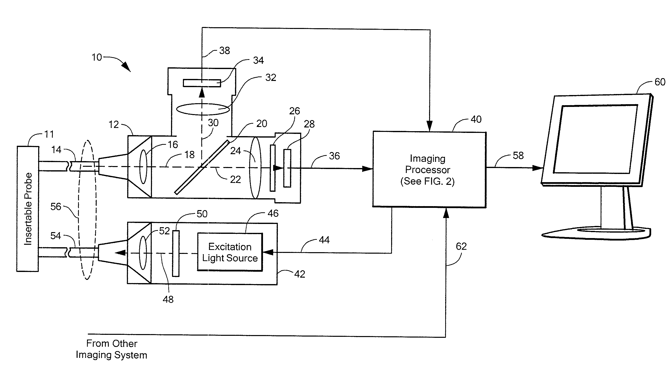
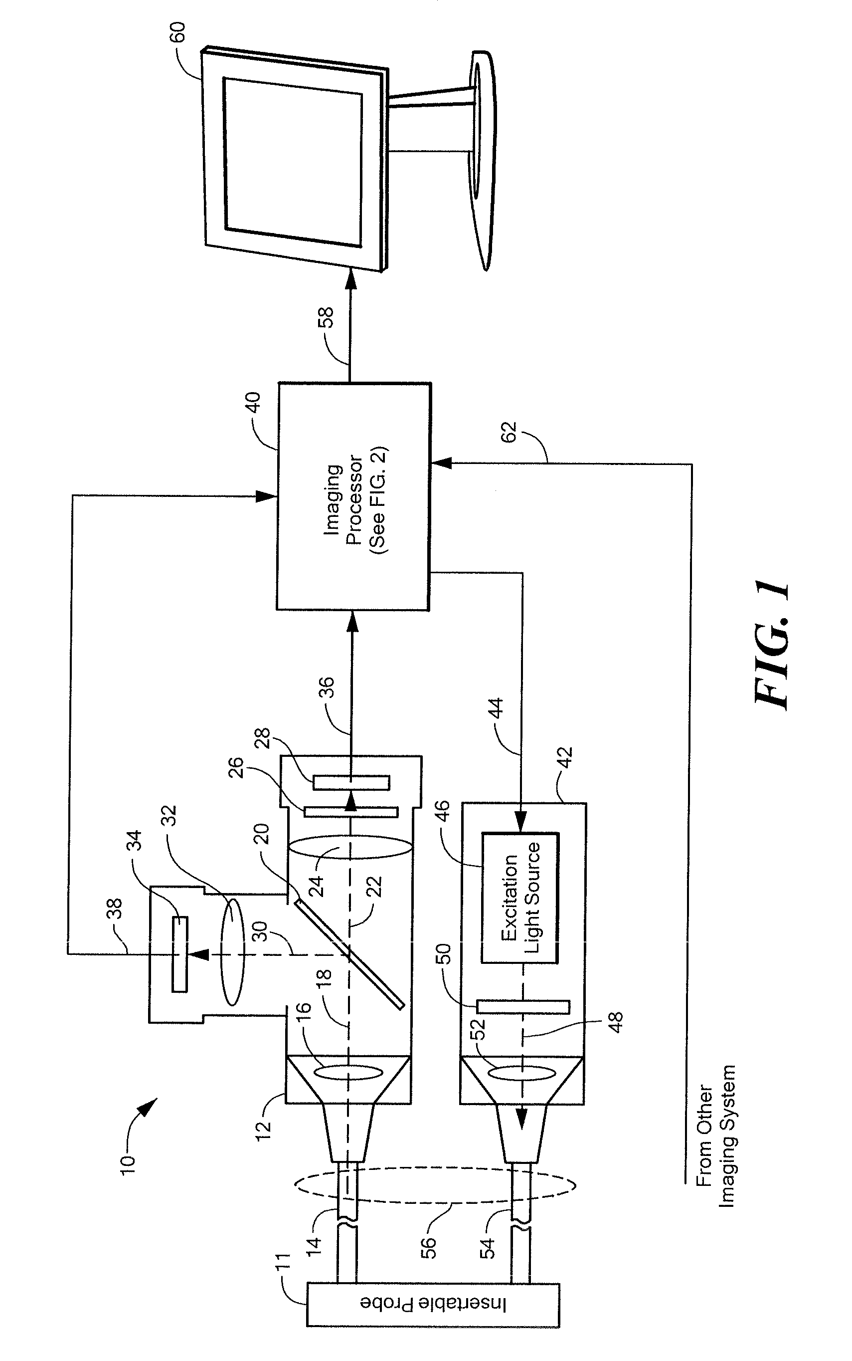
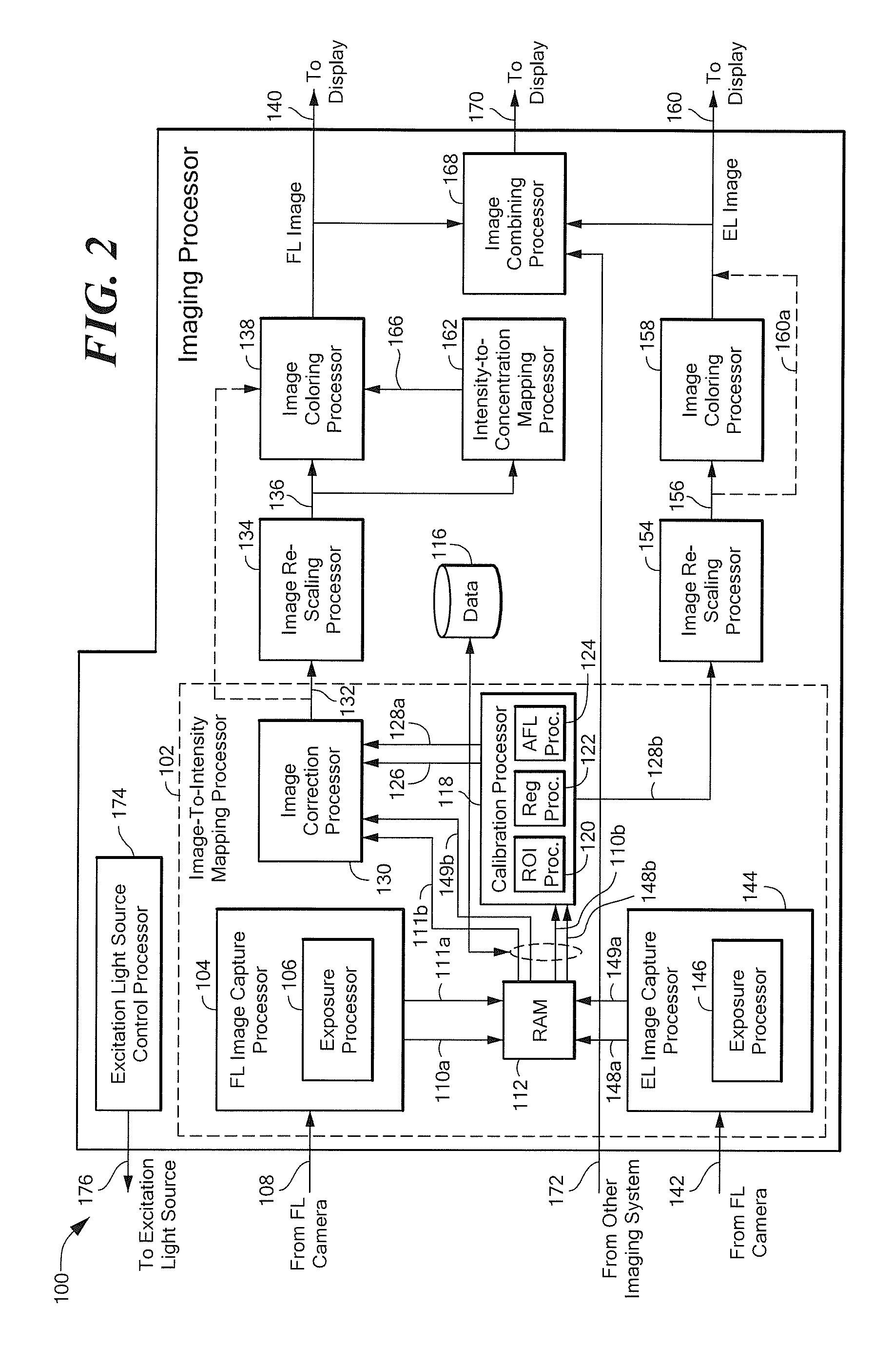
[0050]Before describing the present invention, some introductory concepts and terminology are explained. As used herein, the term “excitation light” is used to describe light generated by an excitation light source. The excitation light includes, but is not limited to, spectral light components (i.e., wavelengths) capable of exciting fluorescence from a biological tissue. The spectral components in the excitation light that are capable of exciting fluorescent light can include a single wavelength, a single band of wavelengths, more than one wavelength, or more than one spectral band of wavelengths. The spectral components in the excitation light that are capable of exciting fluorescent light can include one or more wavelengths in the visible spectral regions of about 400 to 700 nanometers. However, the spectral components in the excitation light that are capable of exciting fluorescent light can also include one or more wavelengths in the other spectral regions, for example, in the ...


PUM


Abstract

An imaging system divides image pixels intensities by exposure time to generate image data in units of intensity per time. The imaging system divides a fluorescent light image in intensity per time units by an excitation light image in intensity per time units to provide a quantitative corrected fluorescent light image that is generally invariant to position of an imaging instrument relative to a biological tissue being imaged.

Description

STATEMENT REGARDING FEDERALLY SPONSORED RESEARCH[0001]This invention was made with government support under Contract No. RO1-EB001872 awarded by the National Institute of Health. The government has certain rights in the invention.CROSS REFERENCE TO RELATED APPLICATIONS[0002]Not Applicable.FIELD OF THE INVENTION[0003]This invention relates generally to imaging systems and methods and, more particularly, to optical imaging systems and methods adapted to provide fluorescent light images of biological tissue.BACKGROUND OF THE INVENTION[0004]Conventionally, fluorescent light has been used to generate microscopic images of histological slices of biological tissue using so-called fluorescence microscopy. However, tissue sectioning used in conventional fluorescence microscopy is limited to slice thicknesses (i.e., tissue depths) on the order of half a millimeter, and therefore, conventional fluorescence microscopy is not appropriate for imaging through entire organs or through the whole hum...

Claims


Application Information

Patent Timeline
IPC IPC(8): A61B6/00
CPCA61B1/00009A61B1/045A61B1/043A61B1/000094
Inventor MAHMOOD, UMARWEISSLEDER, RALPHSHETH, RAHUL A.UPADHYAY, RABI
Owner THE GENERAL HOSPITAL CORP
Why Eureka
Try Eureka