Methods and Apparatus for Simulaton of Endovascular and Endoluminal Procedures

a technology of endovascular and endoluminal procedures, applied in the field of medical training, can solve the problems of putting patients at risk, challenging to meet training requirements, and the tubular structure itself is not visible in x-ray

Inactive Publication Date: 2008-01-24
THE GENERAL HOSPITAL CORP
View PDF2 Cites 204 Cited by
  • Summary
  • Abstract
  • Description
  • Claims
  • Application Information

AI Technical Summary

Problems solved by technology

Unfortunately, the tubular structures themselves are not visible in X-ray images.
Mistakes naturally occur during this learning process putting patients at risk.
It is believed that 1) there is a need for specialty-specific training, 2) competency is directly related to the number of interventions performed, and 3) it is very challenging to meet the training requirements while at the same time protecting patients from untrained practitioners.

Method used


Image

  • Methods and Apparatus for Simulaton of Endovascular and Endoluminal Procedures
  • Methods and Apparatus for Simulaton of Endovascular and Endoluminal Procedures
  • Methods and Apparatus for Simulaton of Endovascular and Endoluminal Procedures

Examples


Embodiment Construction

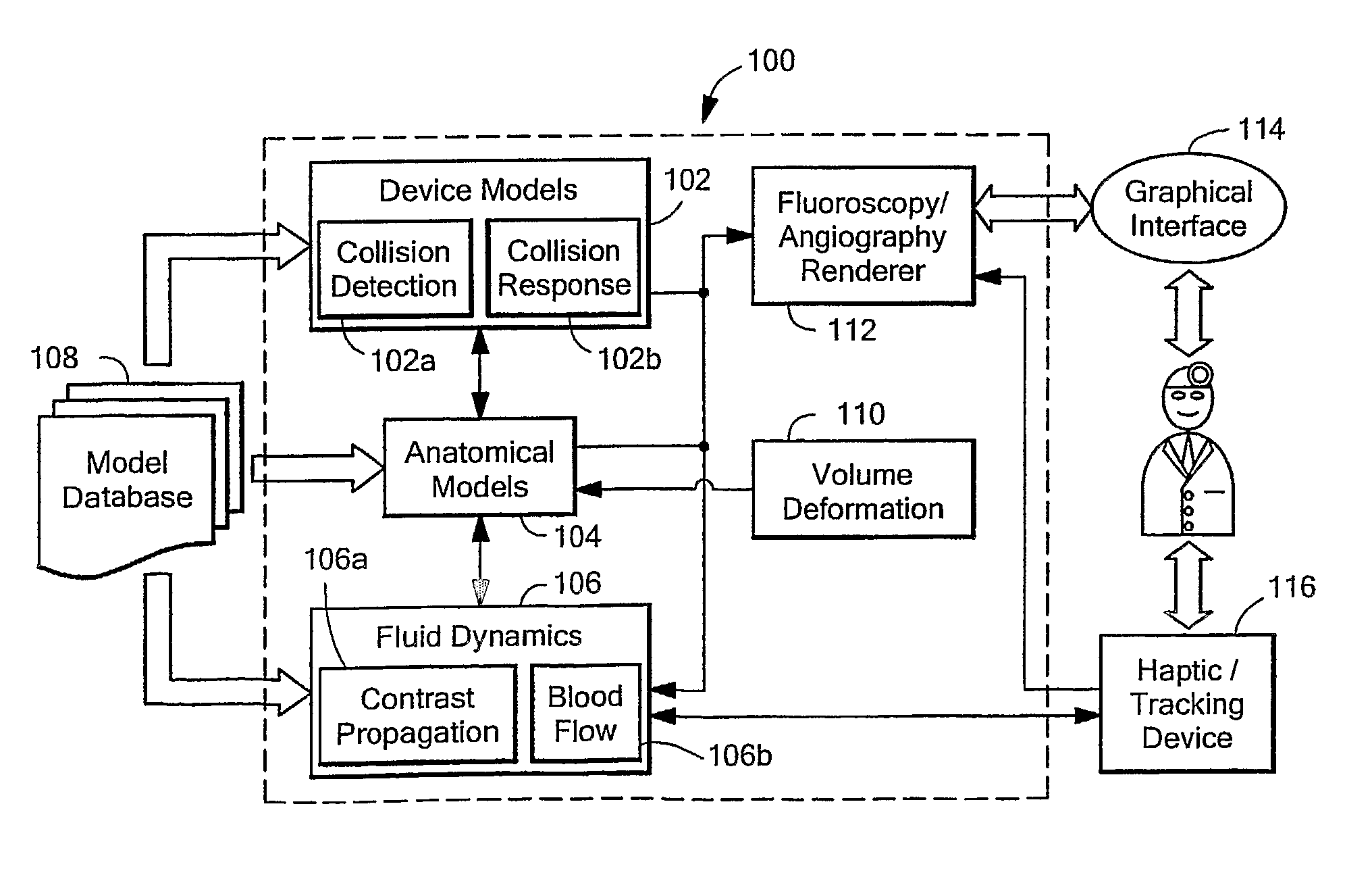
[0049]FIG. 2 is a block diagram of an exemplary interventional radiology procedure training system 100 in accordance with the present invention. The system includes a device model module 102 having a collision detection module 102a and collision response module 102b for a simulated procedure. An anatomical model module 104 models patient anatomy. And a fluid dynamics module 106 includes a contrast propagation module 106a and blood flow module 106b. A model database 108 can provide information to the device model module 102, anatomical model module 104, and fluid dynamics module 106. Volume deformation module 110 can provide information to the anatomical model module 104 and a fluoroscopy / angiography renderer 112 can provide information needed to display information to a user.

[0050] A graphical interface 114 can be provided to enable a user to interact with the system and a haptic / tracking device 116 for various instruments can be coupled to the system, as described more fully below...


PUM


Abstract

Methods and apparatus provide realistic training in endovascular and endoluminal procedures. One embodiment includes modeling accurately the tubular anatomy of a patient to enable optimized simulation. One embodiment includes simulating the interaction between a flexible device and the anatomy and optimizing the computation. One embodiment includes replicating the functionality of therapeutic devices, e.g. stents, and simulating their interaction with anatomy. One embodiment includes computing hemodynamics inside the vascular model. One embodiment includes reproducing visual feedback, using synthetic X-ray imaging and/or or visible light rendering. One embodiment includes generating contrast agent injection and propagation through a tubular network. One embodiment includes reproducing aspects of the physical environment of an operating room by simulating or tracking, such as C-arm control panel, foot pedals, monitors, real catheters and guidewires, etc. One embodiment includes tracking instrument position and mimicking haptic feedback experienced when manipulating certain medical devices.

Description

FIELD OF THE INVENTION [0001] The present invention relates to medical training and, more particularly, to devices and systems for providing realistic training in endovascular and endoluminal procedures. BACKGROUND OF THE INVENTION [0002] As is well known in the art, over the last thirty years medicine has been revolutionized by the advent of minimally invasive techniques to treat disease without the need for surgery. Among the most widely practiced of these new minimally invasive procedures are interventional vascular and visceral procedures and flexible endoscopy. These interventional procedures such as balloon dilatation of strictures, stenting, and catheter-based drug delivery have substantially improved the outcomes for patients with various diseases. [0003] In flexible endoscopy, entry is made through a natural orifice or a small surgical incision. Interventional fluoroscopic procedures are initiated via a percutaneous puncture in which a guidewire-catheter combination is inse...

Claims


Application Information

Patent Timeline
Patent Type & Authority Applications(United States)
IPC IPC(8): G09B23/30
CPCG09B23/285G16H50/50
Inventor COTIN, STEPHANE M.WU, XUNLEINEUMANN, PAUL F.LENOIR, JULIEN R.DURIEZ, CHRISTIAN M.BRADSLEY, RAYN S.PEGORARO, VINCENTDAWSON, STEVEN M.
Owner THE GENERAL HOSPITAL CORP
Why Eureka
Try Eureka