Method and apparatus for tissue resection

a tissue resection and tissue technology, applied in the field of tissue resection devices and methods, can solve the problems of increasing the complexity, time and expense of the resection procedure, affecting the effect of hemostasis promotion, and severing the tissue, so as to prevent the polymerization of the hemostasis-promoting material

Inactive Publication Date: 2008-01-24
BOSTON SCI SCIMED INC
View PDF4 Cites 772 Cited by
  • Summary
  • Abstract
  • Description
  • Claims
  • Application Information

AI Technical Summary

Benefits of technology

[0023]In still another aspect of the invention, a resection device includes an elongated probe shaft and a tissue resection member disposed at a distal end of the elongated probe shaft. The tissue resection member includes a cutting surface configured for being placed into contact with tissue. A porous delivery member is disposed at the distal end of the elongated probe shaft and is coupled to a source of polymerizable hemostasis-promoting material. An ultra violet light emitter is located adjacent to the porous delivery member. In order to prevent polymerization of the hemostasis-promoting material within porous delivery member, a light shield is interposed between the porous delivery member and the ultra violet light emitter.
[0024]In yet another aspect of the invention, a method of resecting tissue includes providing a resection device of the type described above. Using the probe, the tissue is cut with the cutting surface of the tissue resection member. In addition, the polymerizable hemostasis

Problems solved by technology

When used to cut tissue, the electrical arcing and corresponding current flow results in a highly intense, but localized heating, sufficient enough to break intercellular bonds, cellular membranes, and cellular contents, resulting in tissue severance.
Regardless of which modality is used, however, extensive bleeding can occur, which can obstruct the surgeon's view and lead to dangerous blood loss levels, requiring transfusion of blood, which increases the complexity, time, and expense of the resection procedure.
In the case where an organ is resected, application of RF energy divides the parenchyma, thereby skeletonizing the organ, i.e., leaving vascular tissue that is typically more difficult to cut or dissect relative to the parenchyma.
When electrosurgically resecting tissue, care must be taken to prevent the heat generated by the electrode from charring the tissue, which generates an undesirable odor, results in tissue becoming stuck on the electrosurgical probe, and most importantly, increases tissue resistance, thereby reducing the efficiency of the procedure.
As a result, if the fluid and removed tissue is not effectively aspirated from the tissue site, the elec

Method used


Image

  • Method and apparatus for tissue resection
  • Method and apparatus for tissue resection
  • Method and apparatus for tissue resection

Examples


Embodiment Construction

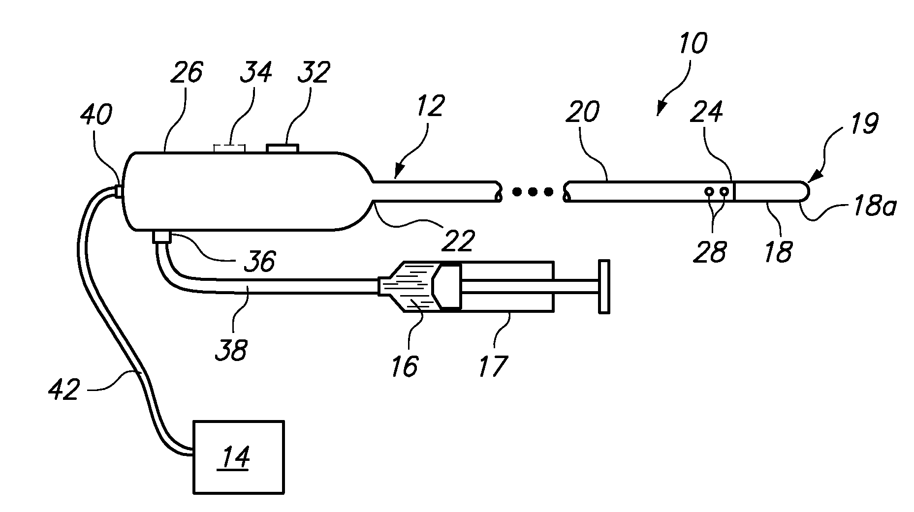
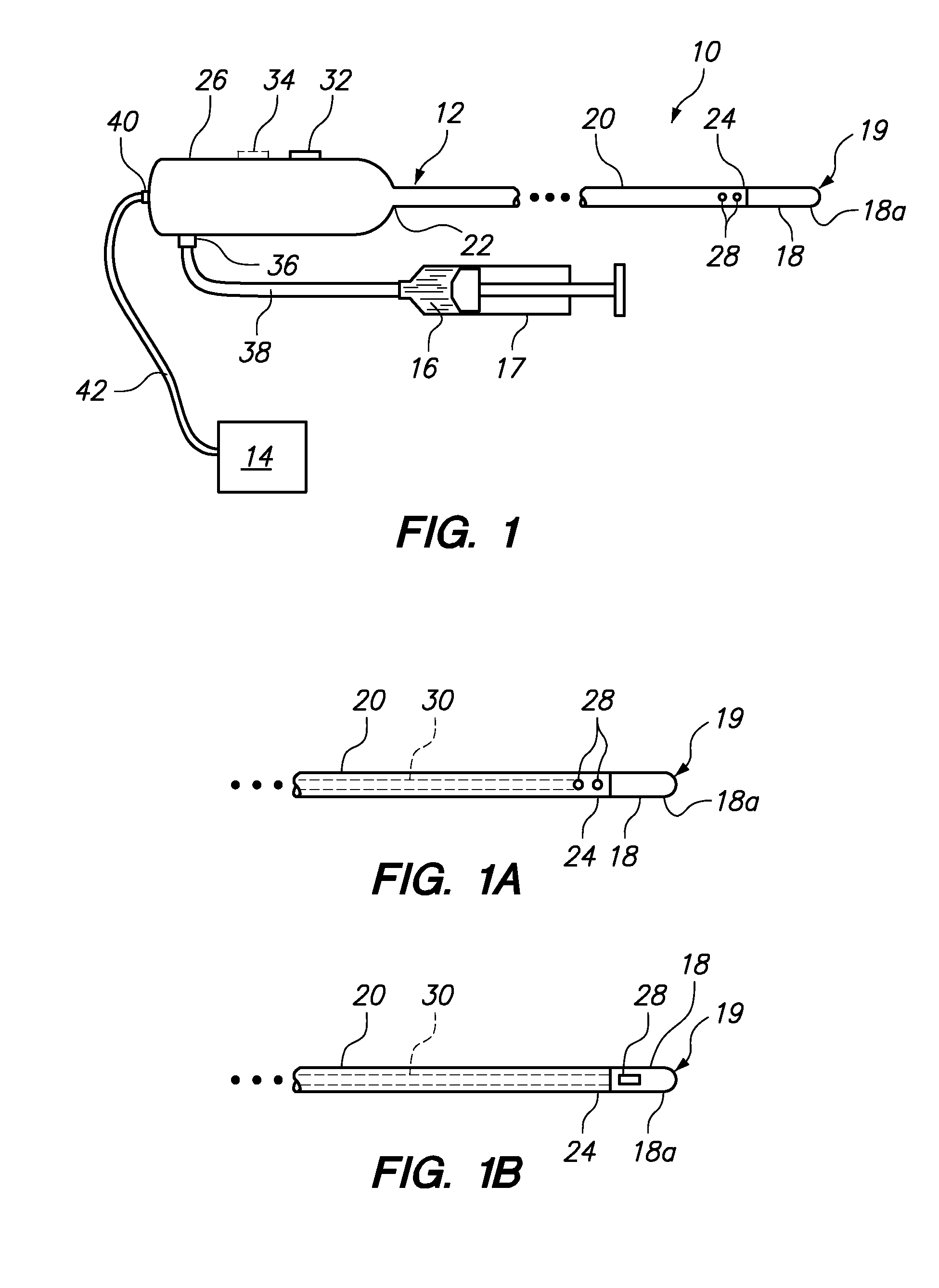
[0049]FIG. 1 illustrates a tissue resection device 10 constructed in accordance with one embodiment of the present invention. In this embodiment, the tissue resection device 10 generally includes a resection device or probe 12 configured for resecting and coagulating tissue and a syringe 17 or other pumping source configured for delivering a polymerizable hemostasis-promoting material 16 to the resection site. The resection probe 12 includes a tissue resection member 18 disposed on a distal end of the resection probe 12 that is configured for being placed into contact with tissue.

[0050]In the embodiment illustrated in FIG. 1, tissue resection member 18 is formed as a resection electrode 19. Accordingly, the device 10 further includes an ablation energy source, and in particular a radio frequency (RF) or microwave generator 14, configured for supplying RF energy or microwave energy to the resection electrode 19 in a controlled manner.

[0051]The resection probe 12 generally comprises a...


PUM


Abstract

A resection device includes an elongated probe shaft and a tissue resection member disposed at a distal end of the elongated probe shaft. The tissue resection member has a cutting surface configured for being placed in contact with tissue. In one aspect of the invention, at least one ejection port is located adjacent to the cutting surface of the tissue resection member, wherein the at least one ejection port is coupled to a source of a polymerizable hemostasis-promoting material that is delivered to the resection site of interest. In certain embodiments, polymerization of the hemostasis-promoting material may be accelerated by application of heat, radiofrequency energy, or ultra violet light.

Description

RELATED APPLICATION DATA[0001]The present application claims the benefit under 35 U.S.C. § 119 to U.S. provisional patent application Ser. No. 60 / 807,815 filed Jul. 19, 2006. The foregoing application is hereby incorporated by reference into the present application in its entirety.FIELD OF THE INVENTION[0002]The field of the invention generally relates to tissue resection devices and methods. More particularly, the field of the invention pertains to devices and methods for use in resecting tissue such as, for example, diseased organ tissue.BACKGROUND OF THE INVENTION[0003]Electrosurgery is now a widely used surgical method for treating tissue abnormalities. One class of electrosurgical abalation devices are so-called monopolar electrosurgical devices. Typically such ablation devices include an electrosurgical probe having a first or “active” electrode extending from one end. The electrosurgical probe is electrically coupled to an electrosurgical generator, which provides a high freq...

Claims


Application Information

Patent Timeline
IPC IPC(8): A61B17/32
CPCA61B17/00491A61B17/320068A61B2017/00495A61B18/1482A61B18/22A61B17/3211A61B2017/320069A61B18/20
Inventor OYOLA, ARNOLD E.BUISER, MARCIADICARLO, PAUL
Owner BOSTON SCI SCIMED INC
Why Eureka
Try Eureka