Tissue visualization device having multi-segmented frame

a multi-segment, tissue visualization technology, applied in the field of medical devices, can solve the problems of cramped working area created by inflatable balloons, interference with fine positioning of imaging systems, and inability to expand the working area of balloons,

Inactive Publication Date: 2008-03-06
NIDUS MEDICAL
View PDF19 Cites 153 Cited by
  • Summary
  • Abstract
  • Description
  • Claims
  • Application Information

AI Technical Summary

Benefits of technology

[0009] An instrument having a low-profile configuration for delivery into and/or through a body and an expandable assembly may be used for retracting or moving tissue from a working distal end of the assembly by utilizing an expandable frame to create a working theater within the body without the need for additional instrumentation. Such an apparatus provides a platform for minimally invasive visualization and therapeutics treatment to be carried out for a variety of...

Problems solved by technology

However, such imaging balloons have many inherent disadvantages.
For instance, such balloons generally require that the balloon be inflated to a relatively large size which may undesirably displace surrounding tissue and interfere with fine positioning of the imaging system against the tissue.
Moreover, the working area created by such inflatable balloons are generally cramped and limited in size.
Furthermore, inflated balloons may be susceptible to pressure changes in the surrounding fluid.
However, such additional instruments and insufflation of the body introduces additional complications and time into a procedure.
Accordingly, these types of imaging modalities are generally unable to provide desirable images useful for sufficient diagnosis and therapy of the endol...

Method used


Image

  • Tissue visualization device having multi-segmented frame
  • Tissue visualization device having multi-segmented frame
  • Tissue visualization device having multi-segmented frame

Examples


Embodiment Construction

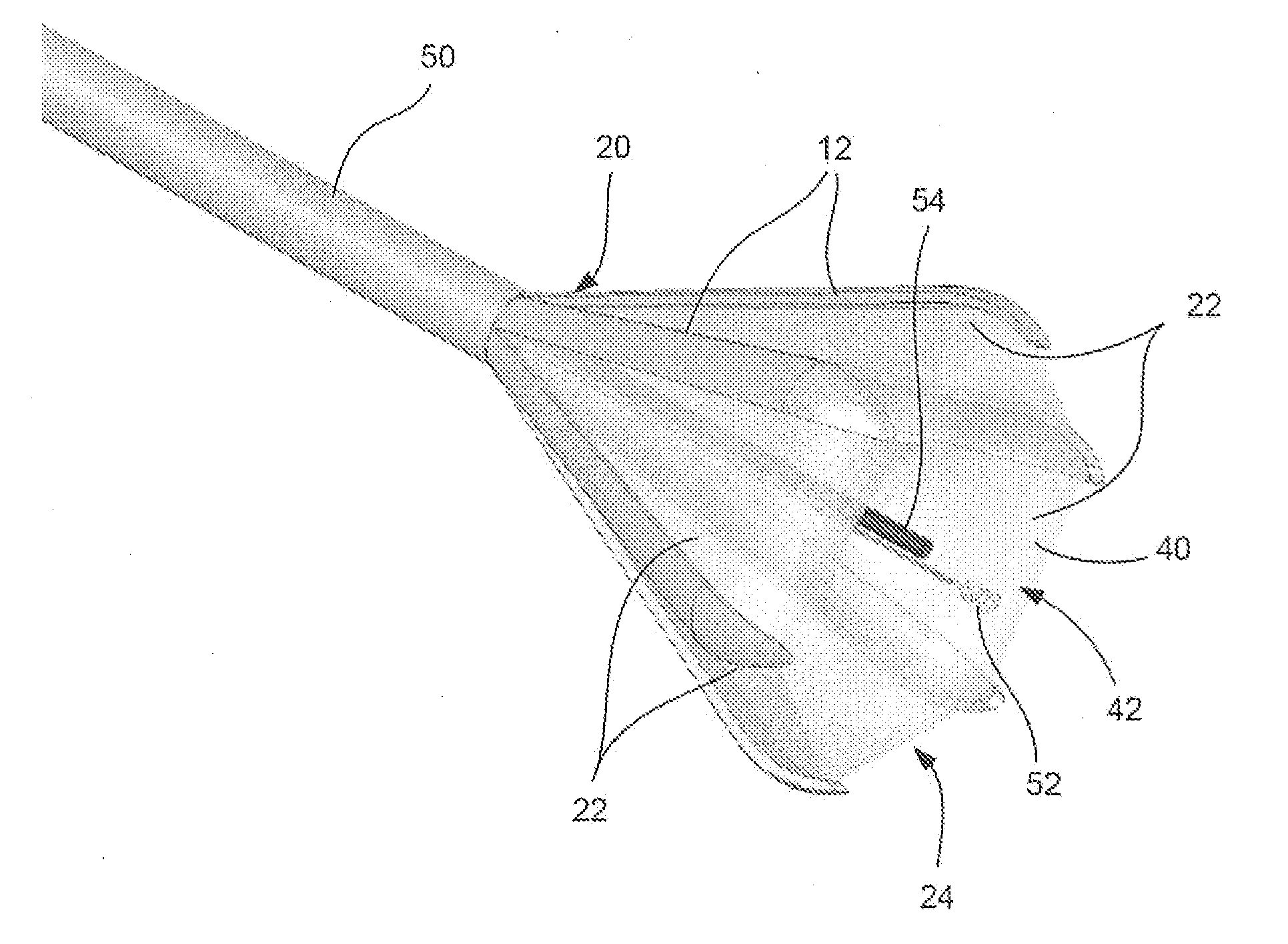
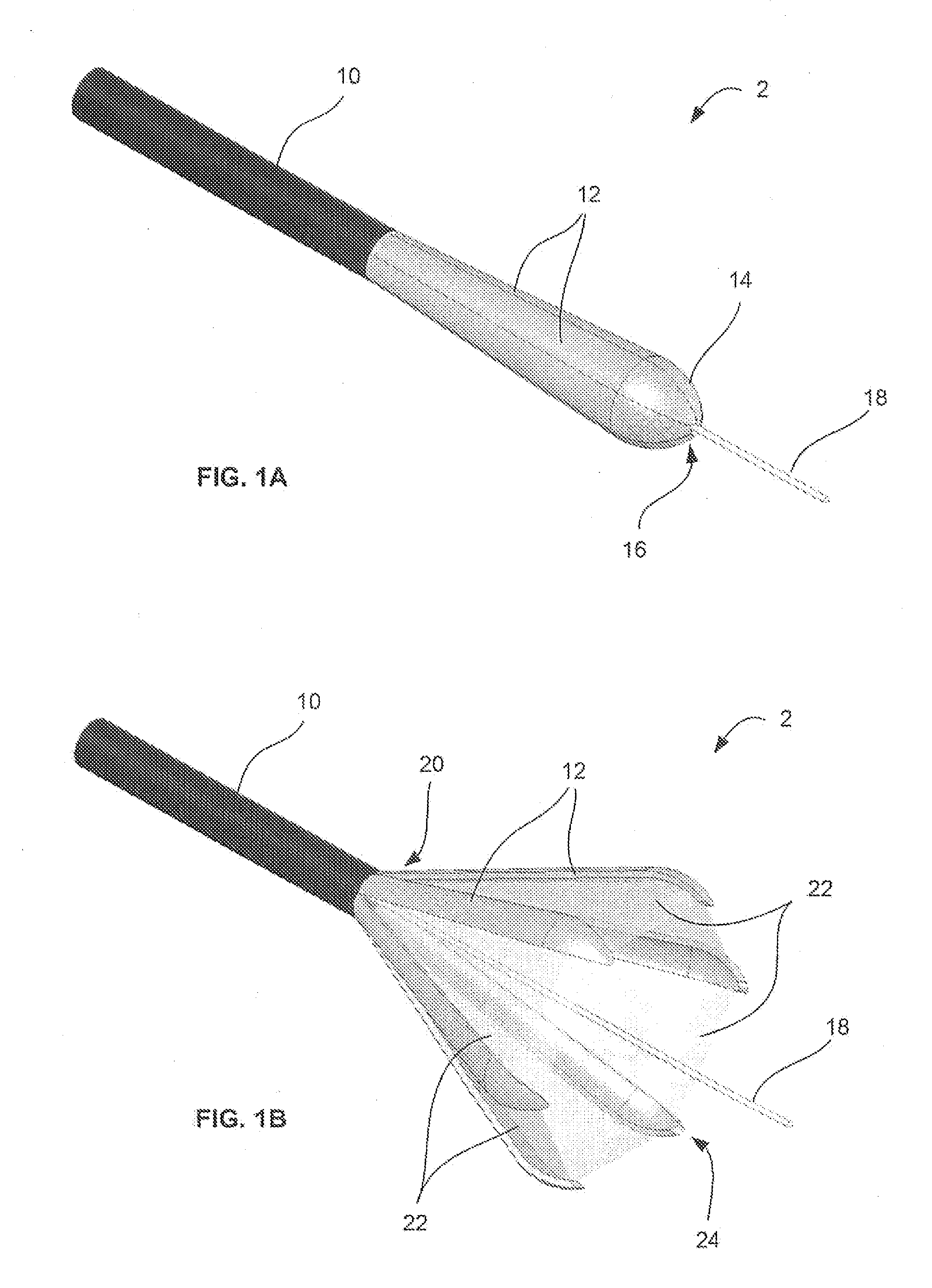
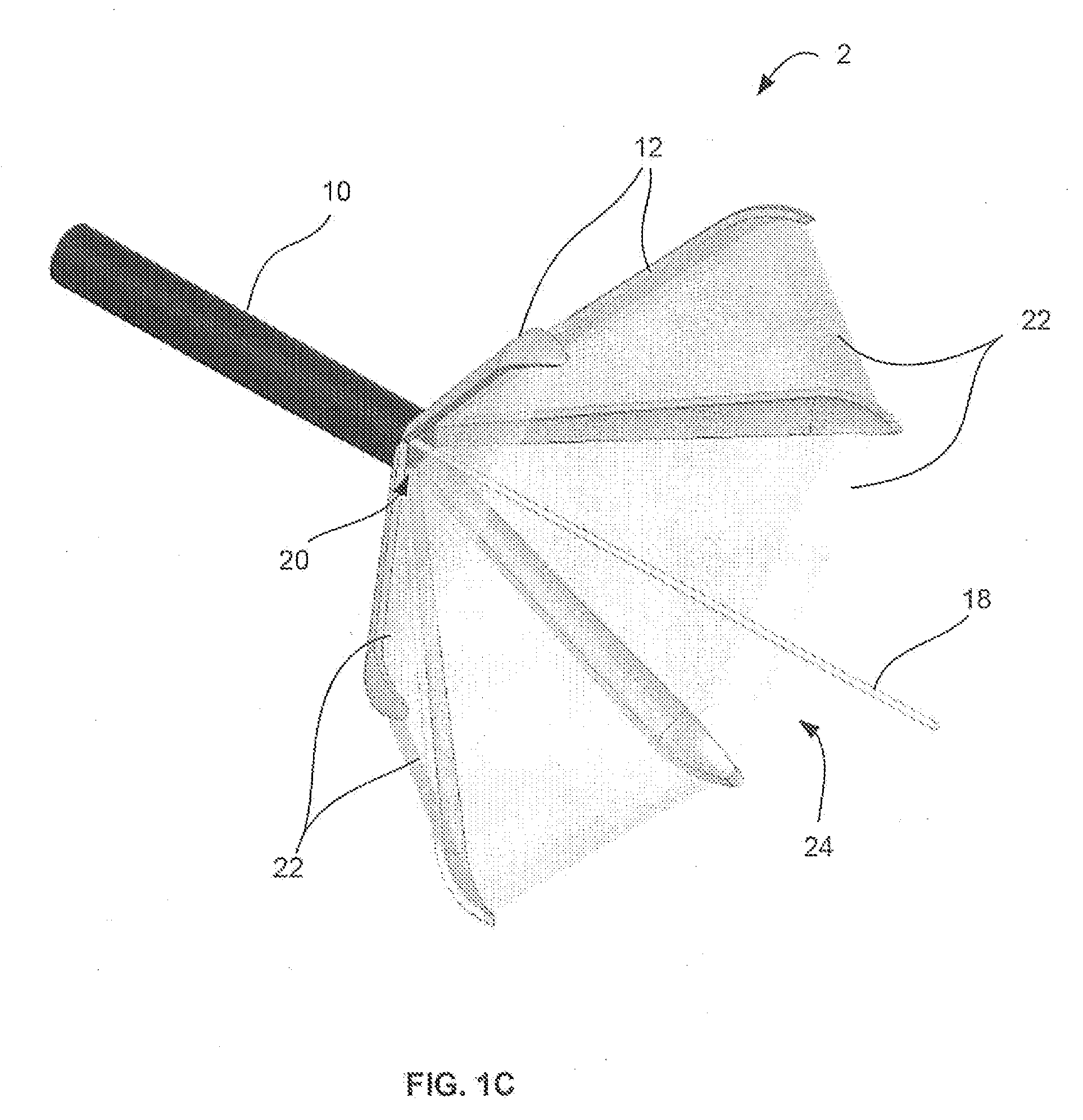
[0026] In performing any number of procedures within a body lumen or body cavity, such as within a heart chamber, peritoneal or thoracic cavity, etc. of a patient, an instrument having a low-profile configuration for delivery into and / or through a body and an expandable assembly for retracting or moving tissue from a working distal end of the assembly may utilize an expandable frame to create a working theater within the body without the need for additional instrumentation. Such an apparatus provides a platform for minimally invasive visualization and therapeutics treatment to be carried out for a variety of procedures in different areas including, but not limited to, e.g., trans-septal access and / or patent foramen ovale closure in cardiac surgery, cutting of the corrugator muscle and accessing the breast from the navel in cosmetic surgery, placing of neuro-stimulator lead for pain management, implanting of artificial disks and injecting of artificial nucleus to the spine, visualiza...


PUM


Abstract

Tissue visualization devices having multi-segmented frame are described herein where such devices may utilize multiple expanding frame members coupled to a flexible deployment catheter shaft or rigid shaft. The multiple frame members may extend distally to collapse into a low-profile configuration and may further expand or open radially to create a working field between the frame members. Moreover, the distal ends of each frame member may be tapered such that the frame members may close tightly relative to one another forming an atraumatic end. Additionally, any number of therapeutic tools can also be passed through the catheter or shaft for performing any number of procedures on the tissue for identifying, locating, and/or treating tissue.

Description

CROSS-REFERENCE TO RELATED APPLICATIONS [0001] This application claims the benefit of priority to U.S. Prov. Pat. App. 60 / 824,417 filed Sep. 1, 2006, which is incorporated herein by reference in its entirety.FIELD OF THE INVENTION [0002] The present invention relates generally to medical devices used for accessing, visualizing, and / or treating regions of tissue within a body. More particularly, the present invention relates to tissue visualization devices having an expandable multi-segmented frame for accessing and / or treating tissue within a patient. BACKGROUND OF THE INVENTION [0003] Conventional devices for accessing and visualizing interior regions of a body lumen are known. For example, ultrasound devices have been used to produce images from within a body in vivo. Ultrasound has been used both with and without contrast agents, which typically enhance ultrasound-derived images. [0004] Other conventional methods have utilized catheters, endoscopes, or probes having position sens...

Claims


Application Information

Patent Timeline
IPC IPC(8): A61B1/32
CPCA61B1/00085A61B1/32A61B1/041A61B1/005
Inventor SAADAT, VAHIDPEH, RUEY-FENGTAM, EDMUND A.
Owner NIDUS MEDICAL
Why Eureka
Try Eureka