User interface for efficiently displaying relevant oct imaging data

Active Publication Date: 2008-05-01
CARL ZIESS MEDITEC INC
View PDF17 Cites 506 Cited by
  • Summary
  • Abstract
  • Description
  • Claims
  • Application Information

AI Technical Summary

Benefits of technology

[0025] The analysis of the change over time of physical attributes is a well-known diagnostic tool. Herein is p

Problems solved by technology

Large medical imaging data sets, such as those acquired during volumetric imaging, present difficulties in displaying relevant information to operators/users.
However, selection of these images can be time-consuming and there is a need to improve the means for accessing relevant slices.
However, the terms are often used interchangeably in the literature and the distinction is often not relevant, since a slice could have been a B-scan under an alternative scanning sequence.
Loss of vis

Method used


Image

  • User interface for efficiently displaying relevant oct imaging data
  • User interface for efficiently displaying relevant oct imaging data
  • User interface for efficiently displaying relevant oct imaging data

Examples


Embodiment Construction

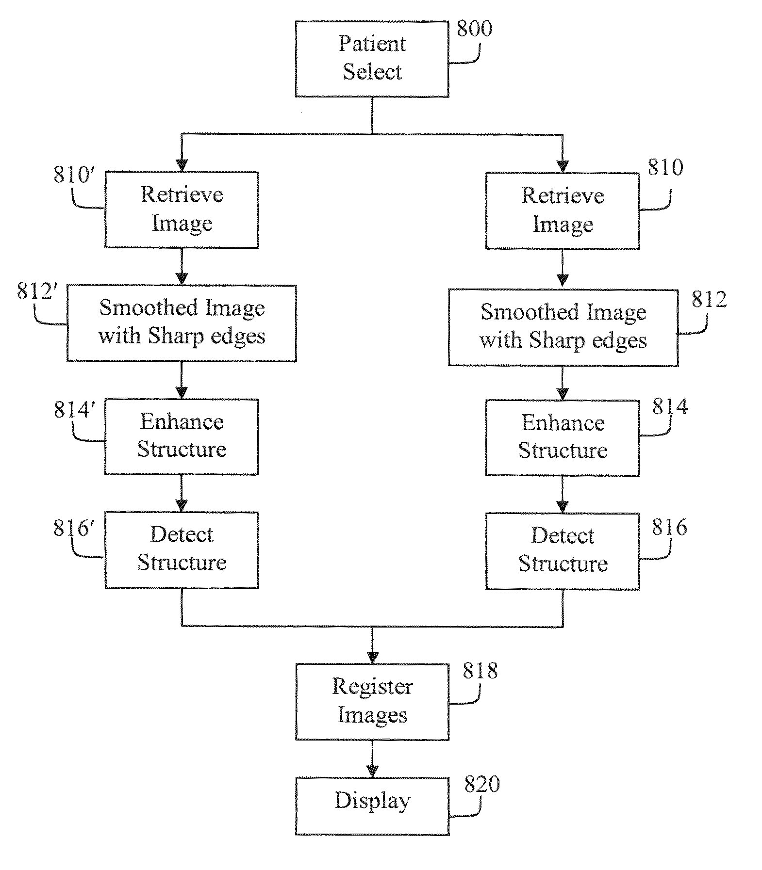
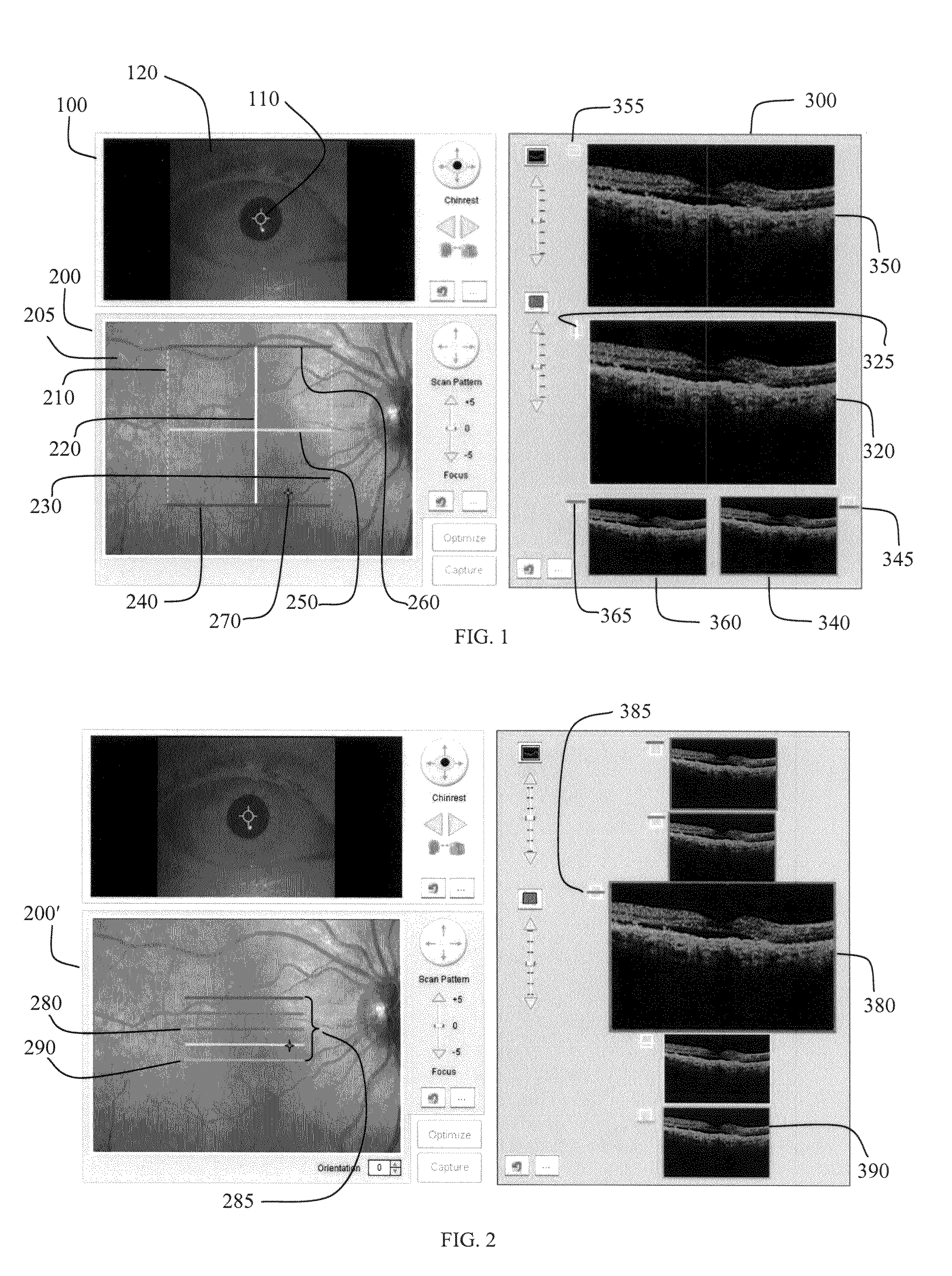
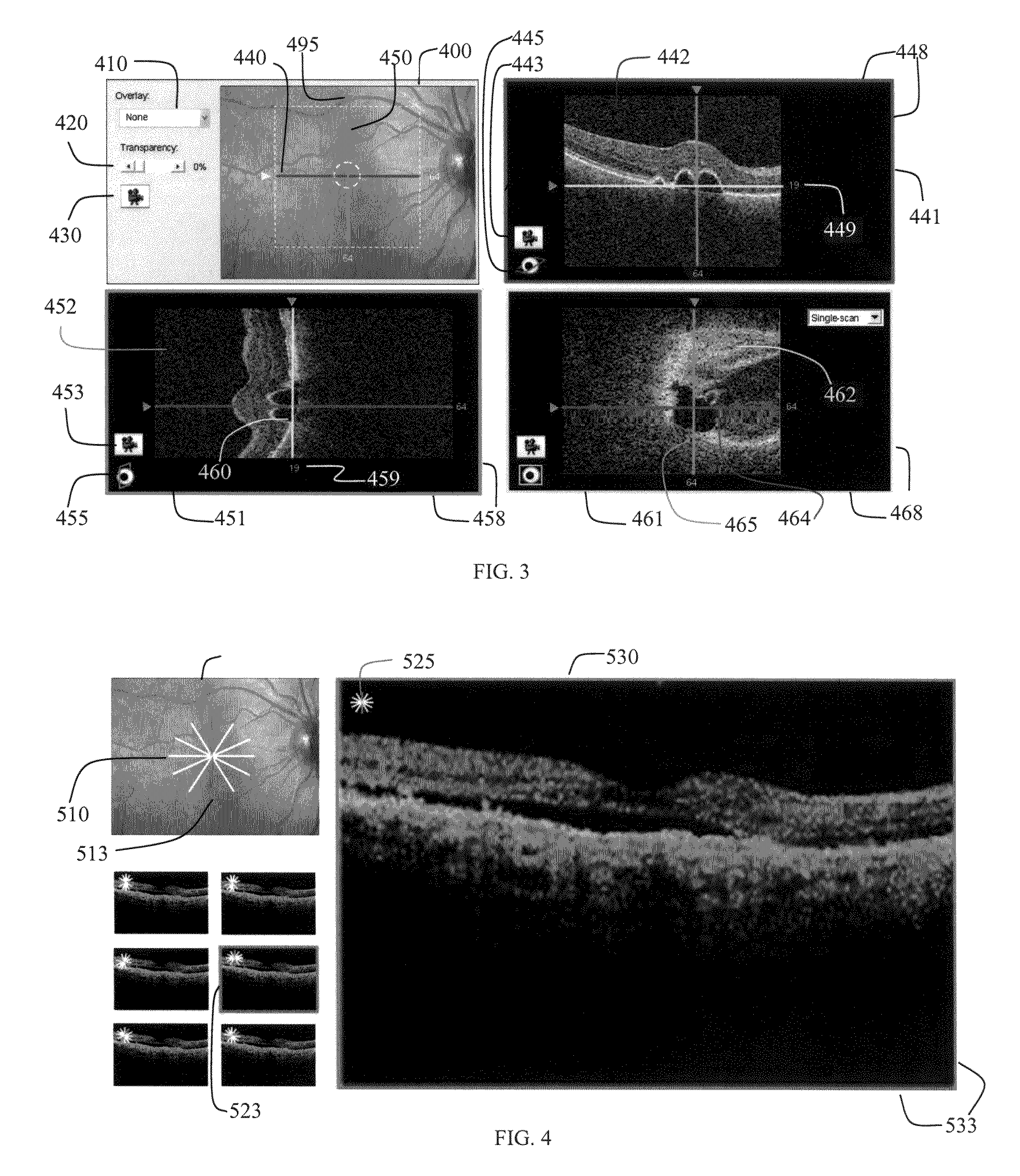
[0043] The present invention is a User Interface (UI) efficiently providing the user with relevant OCT image displays. In one instance, the UI simultaneously displays images of the same region acquired during examinations performed at separate visits. Such displays enable the service provider to monitor changes in the patient's condition over time. The User Interface disclosed is useful for acquiring data, reviewing acquired data, simultaneously viewing multiple images, and manipulating analysis displays. The User Interface provides access to analysis applications that identify regions of interest, reduce the data, and display relevant information in an efficient manner. The User Interface uses image overlays to increase information density in a display area with minimal impact to the underlying display. Overlays help the user find, understand the location of, and visualize relevant data. Image thumbnails and composite image thumbnails are used to readily recognize (and optionally r...


PUM


Abstract

The present invention is an OCT imaging system user interface for efficiently providing relevant image displays to the user. These displays are used during image acquisition to align patients and verify acquisition image quality. During image analysis, these displays indicate positional relationships between displayed data images, automatically display suspicious analysis, automatically display diagnostic data, simultaneously display similar data from multiple visits, improve access to archived data, and provide other improvements for efficient data presentation of relevant information.

Description

PRIORITY [0001] This application claims the benefit of the filing date under 35 U.S.C.§ 119(e) of Provisional U.S. Patent Application Ser. No. 60 / 854,872, filed on Oct. 27, 2006, and Provisional U.S. Patent Application Ser. No. 60 / 857,451, filed on Nov. 7, 2006, which are hereby incorporated by reference in their entirety.TECHNICAL FIELD OF THE INVENTION [0002] The present invention relates to optical medical imaging, and in particular to systems that are adapted to perform Optical Coherence Tomography (“OCT”) for use in diagnosis and monitoring of tissue health. BACKGROUND OF THE INVENTION [0003] Optical Coherence Tomography (OCT) is a technology for performing high-resolution real time optical imaging in situ. OCT herein refers to any of the transverse scanning extensions of one-dimensional optical coherence detection techniques generally derived from optical coherence domain reflectometry (OCDR) or optical frequency domain reflectometry (OFDR). OCT is an optical measurement and i...

Claims


Application Information

Patent Timeline
IPC IPC(8): G06F17/00G16H30/20G16H40/63
CPCA61B3/102G06F19/321G06T15/08G06T2219/028A61B5/7445A61B5/0066A61B5/748G06T19/00G16H30/20G16H40/63G01B9/02091A61B3/0025A61B3/0058G06T2210/41G06T19/003
Inventor DASTMALCHI, SHAHRAM SHAWNSTRAUB, JOCHENOAKLEY, JONATHAN D.STETSON, PAUL F.
Owner CARL ZIESS MEDITEC INC
Why Eureka
Try Eureka