
Detecting and counting bacteria suspended in biological fluids

a technology of biological fluids and bacteria, applied in the field of body fluid assaying, can solve problems such as easy errors in light scattering measurement, and achieve the effects of enhancing the signal to noise and/or signal to clatter ratio, and reducing the level of light reflected

Inactive Publication Date: 2008-05-08
WEICHSELBAUM AMNON +2
View PDF7 Cites 31 Cited by
  • Summary
  • Abstract
  • Description
  • Claims
  • Application Information

AI Technical Summary

Benefits of technology

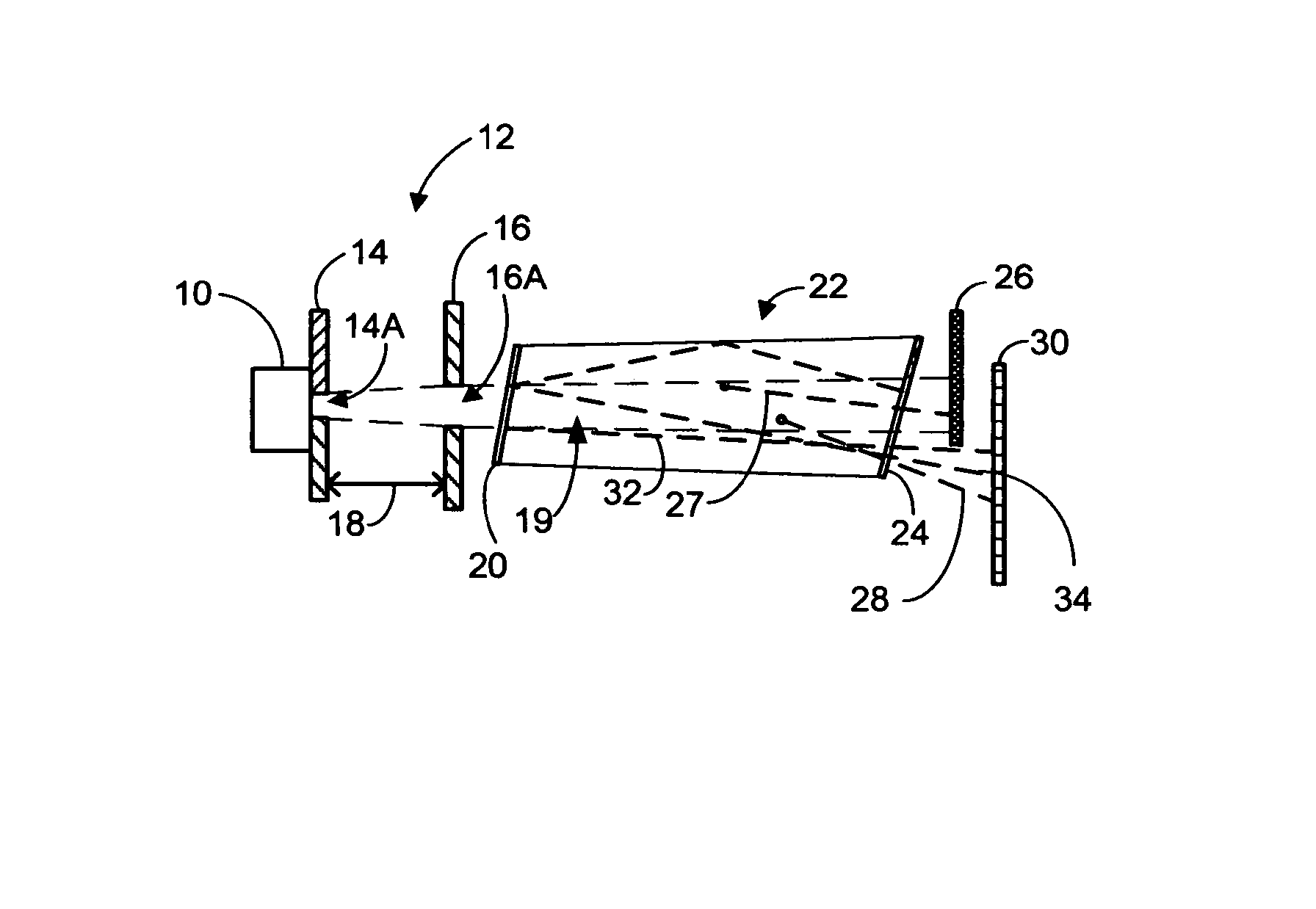
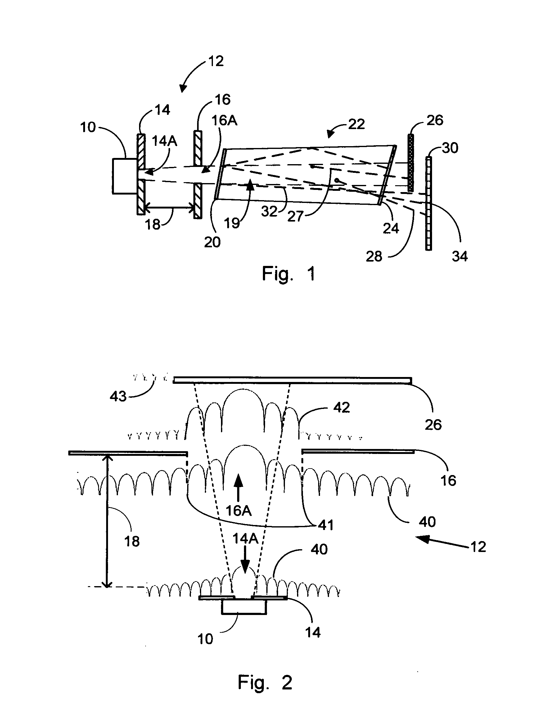
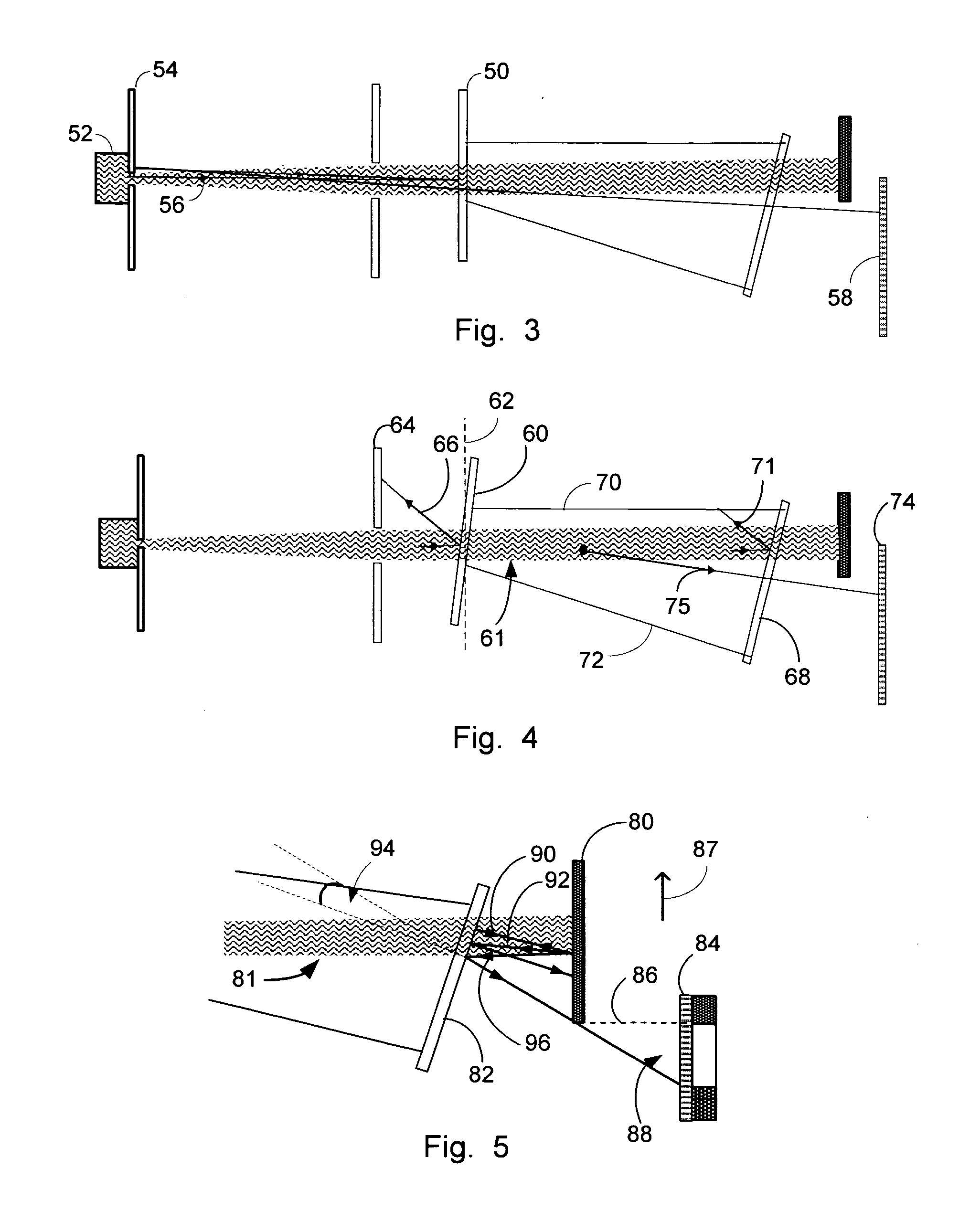
[0007] In accordance with the present invention there is provided a method for detecting bacteria suspended in a biological fluid by spatially filtering the light beam for illuminating the tested fluid, such that the level of the residual illuminating light received at a detecting segment disposed across the surface of the light detector is significantly decreased. Furthermore, in accordance with the present invention there is provided a method for reducing the level of light reflected towards the detecting segment disposed across the surface of the light detector by which the intensity of light scattered by the tested fluid is measured. Such reduction is accomplished by inclining the windows and skewing the sidewall, or sidewalls, of a cuvette adapted to contain a sample of the tested fluid.
[0008] Additionally in accordance with a preferred embodiment of the present invention there is provided a method for substantially canceling out components of the measured speckle images which are stationary in time thereby enhancing the signal to noise and / or signal to clatter ratios of the measured intensities of light scattered by the tested fluid. Scatterers, such as bacteria, which are moving at least in a Brownian motion, induce scattering patterns which are time dependent. Therefore, by successively mapping the instantaneous intensities of light received by the light detector within a predefined exposure time to generate pairs of speckle images; and by respectively subtracting one speckle image of a pair from the other, to form difference plots, most of the features related to the dynamics of the suspended bacteria are retained, whereas the features related to stationary background signals are substantially canceled out.

Problems solved by technology

Light scattering measurements are prone to errors at low concentrations of scatterers, such as of 104 colony forming units per milliliter (CFU / ml) or lower levels.

Method used


Image

  • Detecting and counting bacteria suspended in biological fluids
  • Detecting and counting bacteria suspended in biological fluids
  • Detecting and counting bacteria suspended in biological fluids

Examples


example 1

[0030] In FIGS. 6 and 7 graphs comparing between calculated and measured angular beam profiles generated by means of apertures having different diameters are respectively shown. Plots 100 and 102 respectively correspond to the calculated and measured profiles when employing aperture having a diameter of 150 μm. Similarly plots 110 and 112 respectively correspond to calculated and measured intensities when employing aperture of 500 μm. The intensity of light at a diffraction angle θ I(θ), is given according to diffraction theory by equation (1): I⁡(θ)=(J1⁡(ka⁢ ⁢θ)ka⁢ ⁢θ)2,where(1)

[0031] J1—is the Bessel function of the first kind, a—is the aperture radius, and k—is the wave-number. The calculated and measured intensities shown in a logarithmic scale are measured in arbitrary units (A.U.); the angles are measured in degrees.

[0032] The width of a beam emerging off an aperture of radius a−w(a), is given by equation (2): ω=2⁢a+2π⁢λa⁢L,where(2)

[0033]λ—is the wavelength, a—is the radius...

example 2

[0035] In FIG. 9 a graph comparing between the intensities of the residual illuminating light received at the detector as a function of the scattering angle, with and without the light obscuring means disposed in its place is respectively shown. The intensities shown are measured in arbitrary units (A.U.) employing logarithmic scale. Plot 130 presents the computed beam profile considering the first aperture, which is adjacent to the laser. Dashed line 132 presents the scattering angle corresponding to the diameter of the second apertures which complies with the first zero of the respective Bessel function. Plot 134 presents the intensity of the illuminating beam emerging off the second aperture as a function of the scattering angle when the light obscuring means is avoided. Plot 136 presents the intensity of the residual illuminating light received by the light detector when the light obscuring means is positioned in its place. Dashed line 138 presents the scattering angle correspon...

example 3

[0036] In FIG. 10 a graph comparing among the intensities of the residual illuminating light received at the detector and exemplary scattering profiles computed for various bacterial concentrations is shown. Plot 140 presents the intensity of light received at the light detector as a function of the scattering angle when the light obscuring means is avoided. Plot 144 presents the angular profile of the residual illumination when the light obscuring means is positioned in place. Plots 146, 148, 150, 152 present scattering profiles computed according to the Mie theory for bacterial concentrations of 106, 105, 104, 103 respectively. The bacterial model employed consists of spheres evenly suspended in a liquid, having radii of 1.5 μm whose refracting index equals 1.35, whereas the refracting index of the liquid equals 1.33. For practical purposes and by considering the dynamic ranges of detectors available in the marketplace the maximal scattering angle need not according to the present...


PUM


Abstract

System and method for detecting and counting bacteria suspended in a biological fluid by means of light scattering measurements is provided. In accordance with the method of the invention the level of signal to noise of the measured intensities of light scattered by a sample of the biological fluid is significantly enhanced for forwardly scattered light within a range of scattering angles which are smaller compared to a predefined maximal scattering angle. The system of the invention includes a cuvette adapted to contain a sample of the biological fluid whose sidewalls and windows are suitably constructed and arranged to significantly reduce the level of reflected light obscuring the scattering patterns measured within the range of scattering angles considered.

Description

REFERENCE TO PREVIOUS APPLICATIONS [0001] The present application is a continuation-in-part of U.S. patent application Ser. No. 11 / 573,788, filed on Feb. 16, 2007, which is a .sctn. 371 filing of international patent application Ser. No. PCT / IL05 / 000884, filed on Aug. 16, 2005, claiming the benefit of priority from US application for provisional patent Ser. No. 60 / 601,664, filed on Aug. 16, 2004.FIELD OF THE INVENTION [0002] The present invention relates in general to assaying a body fluid. In particular the present invention relates to optically testing urine, for the presence of bacteria at relatively low bacterial concentrations and to light scattering measurements. BACKGROUND OF THE INVENTION [0003] Detecting and counting bacteria suspended in a biological fluid by means of light scattering measurements is known. In US patent application 20070211251A1, which is incorporated herein by reference, a system and method for detecting and measuring the concentration of bacteria suspend...

Claims


Application Information

Patent Timeline
IPC IPC(8): G01N21/00G01N1/10
CPCG01N21/03G01N21/51G01N2021/513G01N2021/0382G01N2021/4769G01N33/493G01N21/0303G01N2021/479G01N2201/064
Inventor WEICHSELBAUM, AMNONDE LA ZERDA, JAIMEREGEV, ISSAKHAR
Owner WEICHSELBAUM AMNON
Learn More