System and Methods for Inserting a Spinal Disc Device Into an Intervertebral Space
- Summary
- Abstract
- Description
- Claims
- Application Information
AI Technical Summary
Benefits of technology
Problems solved by technology
Method used
Image
Examples
Embodiment Construction
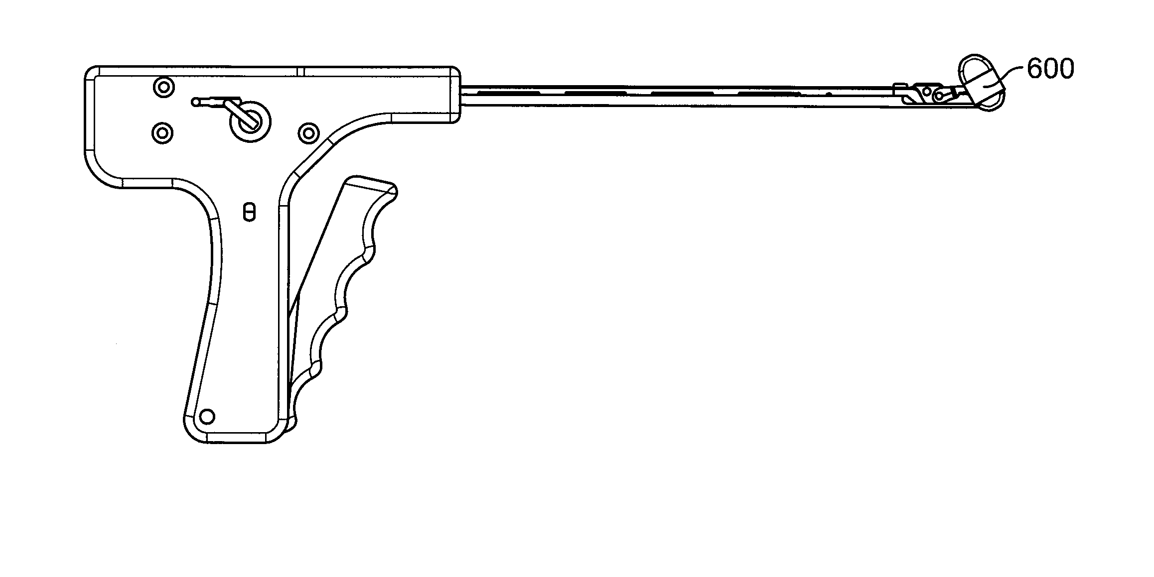
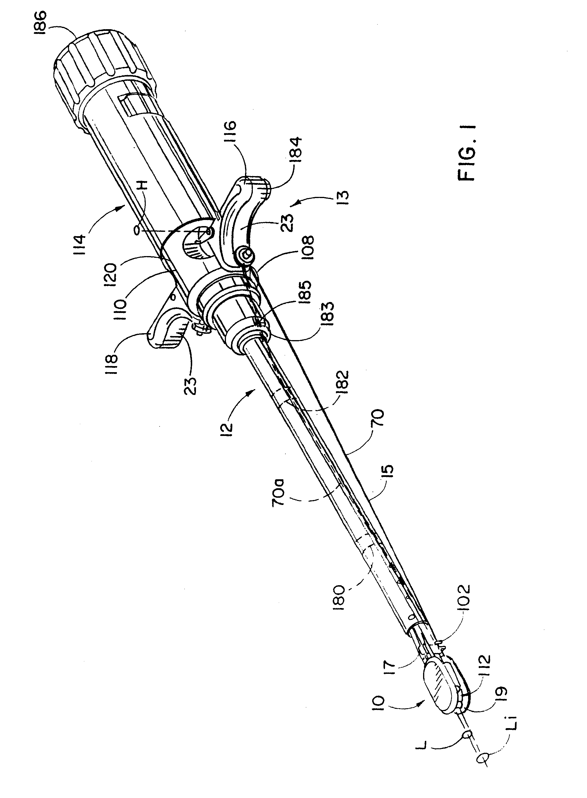
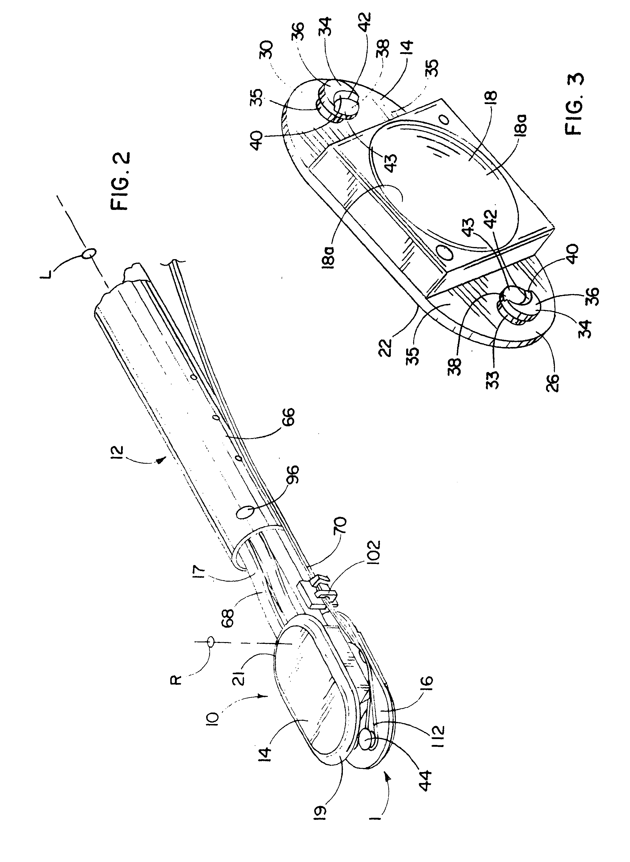
[0183] Referring to FIGS. 1 and 2, a spinal implant 10, such as an artificial disc, is mounted on or connected to an inserter tool 12 for replacing a nucleus of a natural spinal disc between adjacent vertebrae preferably without removing the annulus surrounding the nuclear space. In order to minimize the size of the incision in the annulus, the implant 10 should be inserted by leading with its narrow side facing the incision and pivoting the implant once the implant is positioned within the nuclear space in order to place the implant with its longitudinal dimension or axis Li extending orthogonal to the posterior-anterior direction (i.e. lateral relative to the spine). To pivot the implant 10 without significantly pushing against the incision, which could enlarge or otherwise damage the incision, the inserter 12 has a distal end 17 that extends into the nuclear space and holds the implant 10 while cooperating structure 11 on the inserter 12 and implant 10 are configured to pivot the...
PUM
 Abstract
Description
Claims
Application Information
 - R&D
- Intellectual Property
- Life Sciences
- Materials
- Tech Scout
- Unparalleled Data Quality
- Higher Quality Content
- 60% Fewer Hallucinations
Browse by: Latest US Patents, China's latest patents, Technical Efficacy Thesaurus, Application Domain, Technology Topic, Popular Technical Reports.
© 2025 PatSnap. All rights reserved.Legal|Privacy policy|Modern Slavery Act Transparency Statement|Sitemap|About US| Contact US: help@patsnap.com


