Microscope Viewing Device

a technology for viewing devices and microscopes, applied in the field of microscope viewing devices, can solve the problems of not being able to operate with such devices, not being able to allow simultaneous viewing of data, and not being able to provide such devices

Inactive Publication Date: 2008-05-29
AWDEH RICHARD
View PDF2 Cites 38 Cited by
  • Summary
  • Abstract
  • Description
  • Claims
  • Application Information

AI Technical Summary

Problems solved by technology

While these microscopes are successful in providing a magnified view to the user, they do not allow for the simultaneous view of data, including both real-time streaming data and non-real time data, into the viewer's field (see FIG. 1: the actual microscope view of the surgical field 12, and the ‘black space’10 surrounding the microscopic view).
In the application of surgical microscopes, the utilization of this space in a novel way would allow for real-time intra-operative feedback (including real time and non-real time data, as described in further detail below) to appear in the black space and be viewed by the surgeon in real-time during surgery, but no such devices are currently available.
However, the use of microscopes has historically created a problem for surgeons as their access to, and view of, data while operating is limited to whatever image appears in the viewer's field.
As such, the current technology does not provide surgeons with real time access to parameters that are important to the surgery while they are looking through the surgical microscope.
Further, under the current technology, the surgeon is unable to simultaneously view a magnetic resonance image (MRI), computed tomography image (CT), or any other image or piece of data from a patient's medical record, while he or she is looking through the microscope while operating.
This prevents the surgeon from viewing essential data and from evaluating diagnostic studies while he or she is operating, creating a further limitation with the use of microscopes in surgery and elsewhere.

Method used


Image

  • Microscope Viewing Device
  • Microscope Viewing Device
  • Microscope Viewing Device

Examples


Embodiment Construction

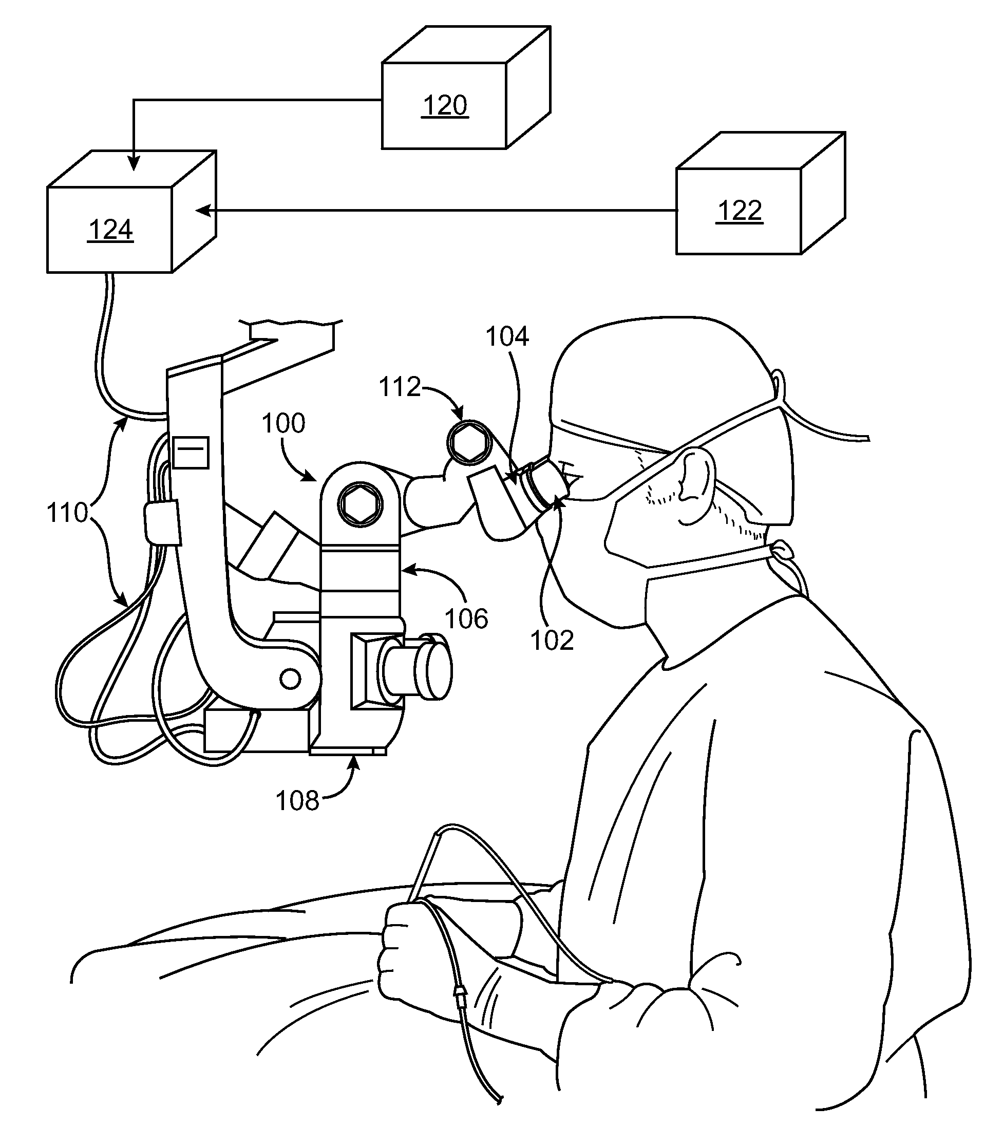
[0029]Referring to the drawing figures, like reference numerals designate identical or corresponding elements throughout the several figures.

[0030]In the context of this application, reference will be made to one or more lightpaths, which indicates the path that light follows from a source, e.g., an object being viewed by a microscope, towards an endpoint. For ease of reference, the term ‘upstream’ will refer to the direction along the lightpath toward the source, and the term ‘downsteam’ will refer to the opposite direction, i.e., along the lightpath away from the source.

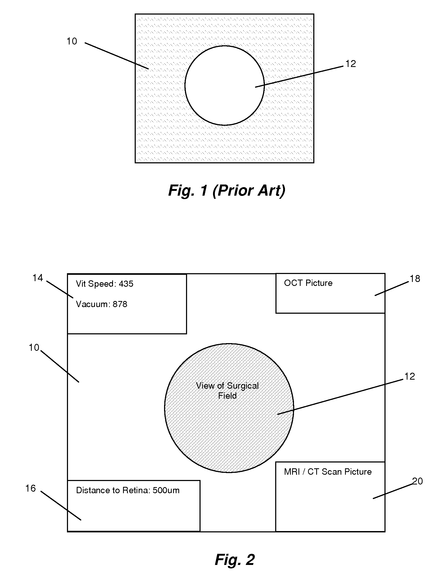
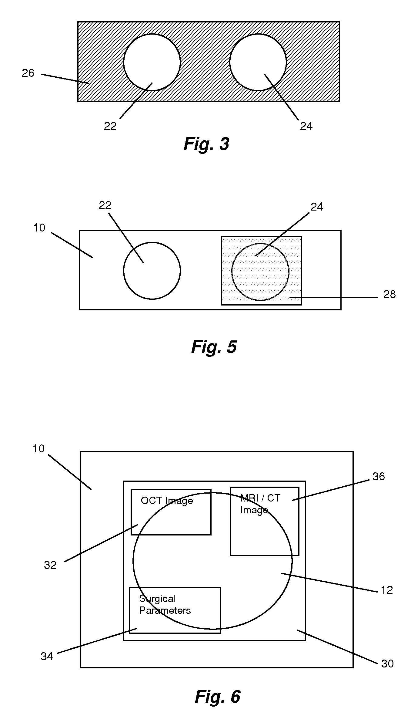
[0031]In general terms, the invention in this application relates to the display of data, graphics, and video, both dynamic and static, inside any microscope such that the user can simultaneously view the microscopic field as well as monitor data parameters. Principles of the present invention can be applied in the setting of any microscope, including surgical microscopes and laboratory microscopes. Through the uti...


PUM


Abstract

A microscope, e.g., a surgical microscope, includes an image generation device in the light path or light paths of the microscope. Images based on real-time or static data concerning the object in view, e.g., a patient's MRI data and / or parameters of one or more surgical instruments being used to operate on the patient, are projected into the microscope's light path so the surgeon does not need to look away from the surgical field and does not require other personnel to read outloud the settings and data from the machines being used in the surgery.

Description

[0001]This application claims priority under 35 U.S.C. § 119 to U.S. Provisional patent application No. 60 / 817,478, filed 30 Jun. 2006, the entirety of which is incorporated by reference herein.BACKGROUND[0002]1. Field of Endeavor[0003]The present invention relates to devices and systems useful as surgical microscopes.[0004]2. Brief Description of the Related Art[0005]Microscopes, including surgical microscopes used in the operating room in human and animal surgery, and laboratory microscopes, offer the user a magnified, stereoscopic view of the selected field. While these microscopes are successful in providing a magnified view to the user, they do not allow for the simultaneous view of data, including both real-time streaming data and non-real time data, into the viewer's field (see FIG. 1: the actual microscope view of the surgical field 12, and the ‘black space’10 surrounding the microscopic view).[0006]Most microscopes include two eyepieces, one for the right eye and one for th...

Claims


Application Information

Patent Timeline
Patent Type & Authority Applications(United States)
IPC IPC(8): G02B21/00
CPCG02B21/368
Inventor AWDEH, RICHARD
Owner AWDEH RICHARD
Why Eureka
Try Eureka