Polymeric joint complex and methods of use

a polymer and joint technology, applied in the field of polymer facet joint complex, can solve the problems of severe limits to the functional ability and quality of life of people, loss of proper alignment or loss of proper articulation of the vertebrae, so as to increase or decrease the constraint of the joint, replace or augment the joint complex, and strengthen the effect of replacing or augmenting the joint complex

Inactive Publication Date: 2008-06-12
GMEDELAWARE 2
View PDF99 Cites 29 Cited by
  • Summary
  • Abstract
  • Description
  • Claims
  • Application Information

AI Technical Summary

Benefits of technology

[0007]One aspect of the invention provides, an implantable device that is placed through a joint space or joint complex, such that a central flexible section reinforces, replaces or augments the joint. The device can be delivered to the joint by access through bone into the joint space without opening or disrupting the joint space. The device reinforces, replaces or augments the joint complex including all or some of the capsule, ligaments, nucleus or other joint complex structures. The flexible central section acts as a flexible and / or conformable spacer with or without providing a fixed axis of rotation. Altering the flexibility of the flexible section can increase or decrease the constraint of the joint. Flexibility can easily be altered or revised in a subsequent procedure after initial implantation.
[0008]Another aspect of the invention provides, devices that allow placement of a device in a joint space without resection or compromising the capsule or surrounding tissue. The devices also allow variable distraction of two bony surfaces. Further the devices and methods do not rely on bony fixation to hold the device in place. However, the devices can use bony fixation, if desired. The devices allow for easy anatomical variations and a wide range of pathologies to be treated as the device contours itself to the surrounding structures. The device also enables a reduction in inventory for hospitals because one size can be adapted to fit many anatomical variations and pathologies.
[0010]In some embodiments, the artificial joint complex the expandable joint segment is adapted to provide a low profile suitable for insertion through an access lumen that accesses a target joint space, such as a minimally invasive lumen formed in the bone. A second, larger profile, is achieved when the expandable segment is inflated while positioned within the lumen of the joint space. In other embodiments, the cannulated anchor is formed integrally with the expandable joint segment, while in still other embodiments, the cannulated anchor is removably connected. A cap for sealing the artificial joint complex is provided to seal the complex once installed and the expandable joint segment has been inflated. In some embodiments, a post, which can be centrally positioned, is positioned within any or all of the cannulated anchor or expandable joint segment. In some embodiments, it is contemplated that the cannulated anchor is a superior cannulated anchor that is adapted to engage a superior articular facet. In other embodiments, the cannulated anchor is an inferior cannulated anchor adapted to engage an inferior articular facet. In either of these embodiments, additional embodiments could provide a second anchor. Where a second anchor is provided, it could be either inferior to the superior cannulated anchor or superior to the inferior cannulated anchor. The second anchor, as with the first anchor, can be cannulated, if desired. Any of the embodiments can provide for the anchors to be threaded, either internally, externally, or both, to achieve the objectives of the design. Alternatively, the anchors could have a smooth exterior surface, a roughened exterior surface, or a coated exterior surface, as desired. The anchors could also be configured to deliver a target agent, such as a pharmaceutical or biological agent.
[0014]In some embodiments, the expandable joint segment is expandable beyond the perimeter of all, or a part, of the joint surfaces. Thus, the expandable joint segment can form a spacer between at least part of the joint surfaces as well as a capsule surrounding at least part of the joint. Additionally, the expandable segment can expand around the joint in such a manner than axial movement of the joint surfaces away from each other is restricted or prevented. The expandable joint segment is typically configured to provide a low profile for insertion through an access lumen, and a larger profile when inflated, such as when it is within the target joint space.

Problems solved by technology

In many cases, the pain severely limits a person's functional ability and quality of life.
Back pain interferes with work, routine daily activities, and recreation.
It is the most common cause of job-related disability and a leading contributor to missed work.
Through disease or injury, the laminae, spinous process, articular processes, facets and / or facet capsule(s) of one or more vertebral bodies along with one or more intervertebral discs can become damaged which can result in a loss of proper alignment or loss of proper articulation of the vertebra.
This damage can result in anatomical changes, loss of mobility, and pain or discomfort.
For example, the vertebral facet joints can be damaged by traumatic injury or as a result of disease.
Diseases damaging the spine and / or facets include osteoarthritis where the cartilage of joint is gradually worn away and the adjacent bone is remodeled, ankylosing spondylolysis (or rheumatoid arthritis) of the spine which can lead to spinal rigidity, and degenerative spondylolisthesis which results in a forward displacement of the lumbar vertebra on the sacrum.
Damage to facet joints of the vertebral body often can also results in pressure on nerves, commonly referred to as “pinched” nerves, or nerve compression or impingement.
The result is pain, misaligned anatomy, and a corresponding loss of mobility.

Method used


Image

  • Polymeric joint complex and methods of use
  • Polymeric joint complex and methods of use
  • Polymeric joint complex and methods of use

Examples


Embodiment Construction

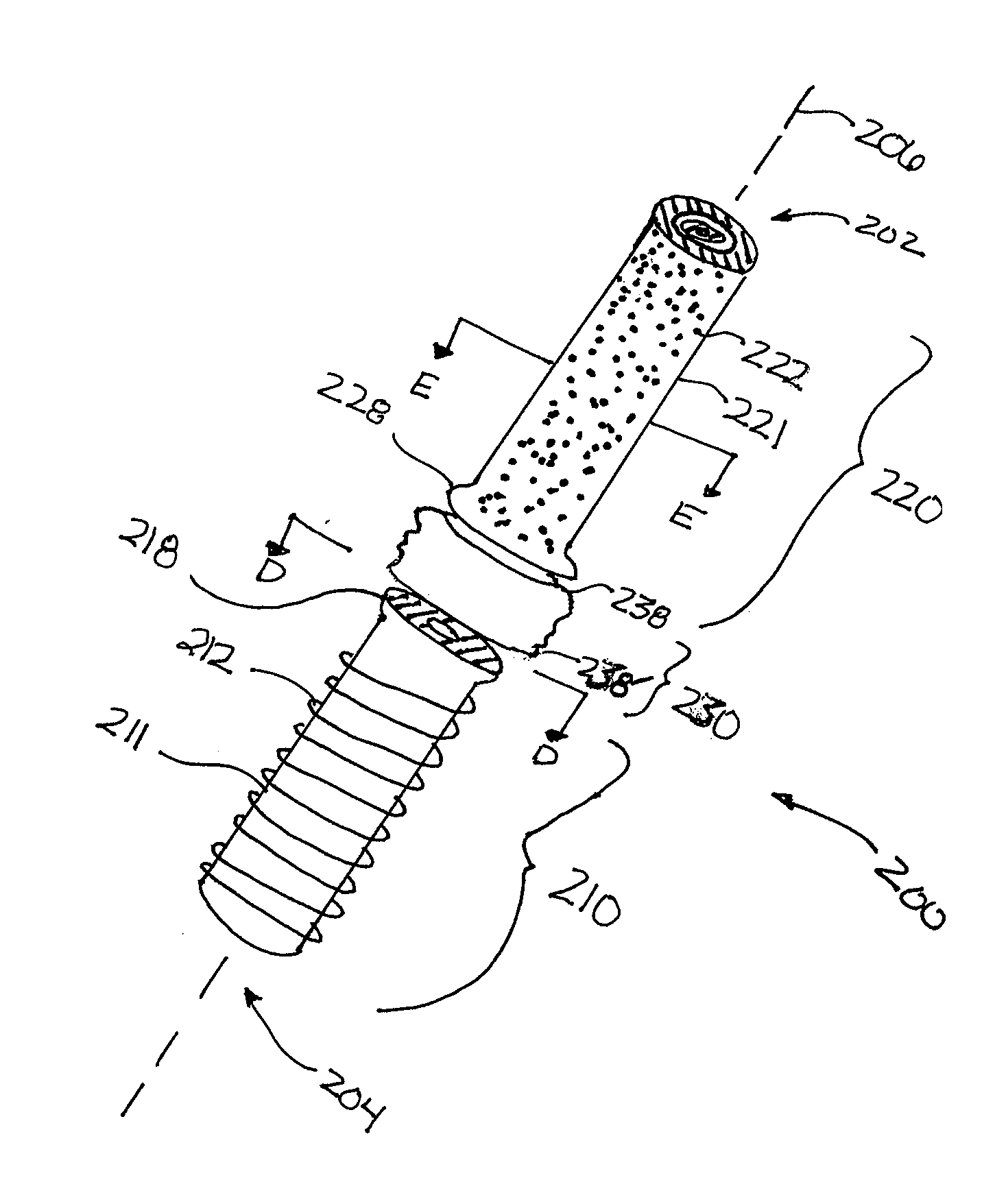
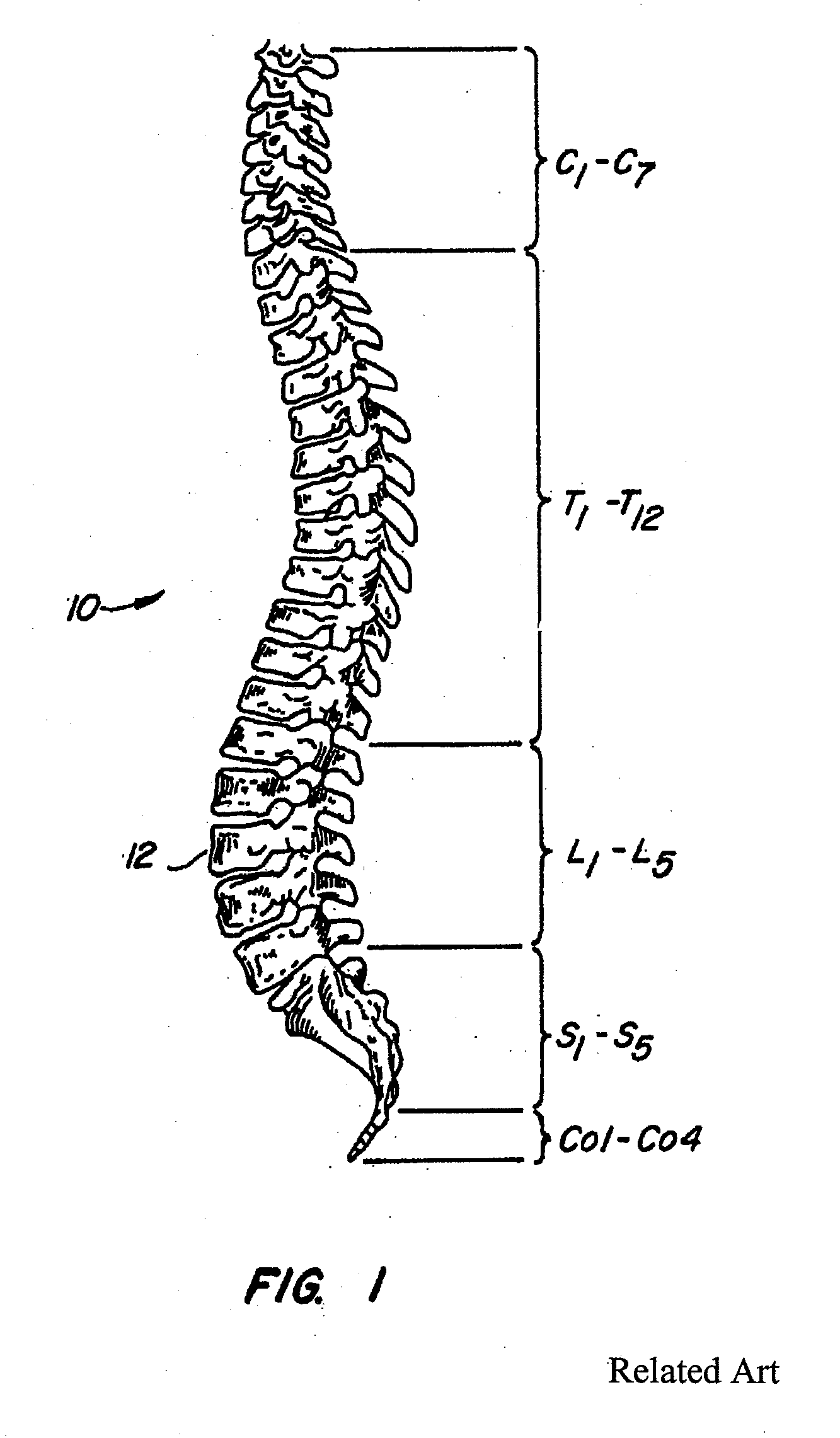
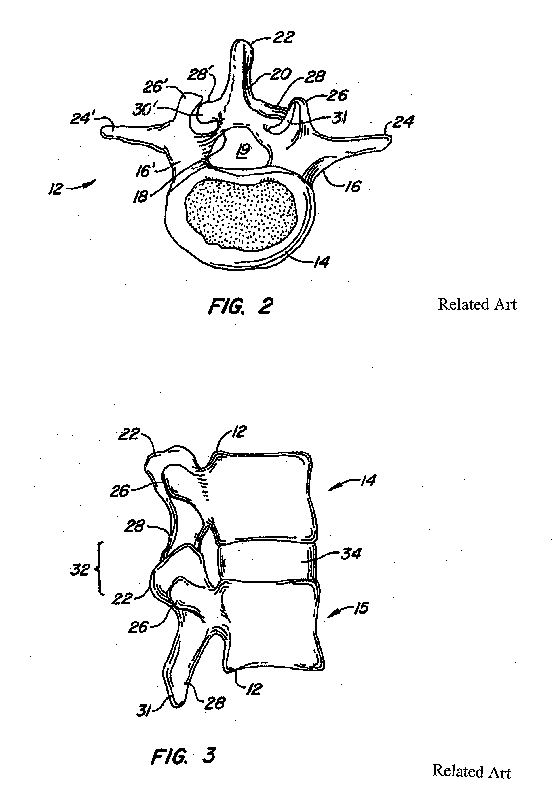
[0046]The invention relates to implantable devices, including implantable prosthesis suitable for implantation within the body to restore, reinforce, replace and / or augment connective tissue such as bone, and systems and methods for treating spinal pathologies. The invention relates generally to implantable devices and apparatuses or mechanisms that are suitable for implantation within a human body to restore, augment, and / or replace soft tissue and connective tissue, including bone and cartilage, and systems for treating spinal pathologies. In various embodiments, the implantable devices can include devices designed to replace missing, removed or resected body parts or structure. The implantable devices, apparatus or mechanisms are configured such that the devices can be formed from parts, elements or components which alone, or in combination, comprise the device. Thus, for example, the implantable devices can be configured such that one or more elements or components are formed in...


PUM

PropertyMeasurementUnit
cylindrical shapeaaaaaaaaaa
shapeaaaaaaaaaa
biocompatibleaaaaaaaaaa

Abstract

The invention describes a variety of implantable artificial joint complexes adapted for implantation within a target joint space within a human body. The joint complexes comprise: an expandable joint segment adapted to fit within the target joint space; and at least one of a first cannulated anchor adapted to engage the expandable joint segment and adapted to engage a bony structure adjacent the target joint space; and a second anchor adapted to engage the expandable joint segment and adapted to engage a bony structure adjacent a target joint space. The invention also discloses methods of implanting a patient specific artificial joint complex. The methods include the steps of: accessing a target joint space by creating an access hole through an adjacent bony structure; inserting a joint complex device having a cannulated anchor and an expandable joint segment through the access hole with the expendable joint segment being positioned between the surfaces forming the joint; injecting material into the expandable joint segment; and sealing access to the target joint space.

Description

CROSS-REFERENCE[0001]This application is a continuation of U.S. patent application Ser. No. 11 / 244,420, filed Oct. 4, 2005, which claims the benefit of U.S. Provisional Patent Application Ser. No. 60 / 616,093, filed Oct. 4, 2004, and entitled “Polymer Joint Complex”, which is also incorporated herein by reference.FIELD OF THE INVENTION[0002]This invention relates to implantable spinal devices, systems, and methods for treating various types of spinal pathologies. The invention relates in particular to a polymeric facet joint complex providing a flexible artificial joint complex.BACKGROUND OF THE INVENTION[0003]Back pain, particularly in the small of the back, or lumbosacral region (L4-S1) of the spine, is a common ailment. In many cases, the pain severely limits a person's functional ability and quality of life. Back pain interferes with work, routine daily activities, and recreation. It is estimated that Americans spend $50 billion each year on low back pain alone. It is the most co...

Claims


Application Information

Patent Timeline
Patent Type & Authority Applications(United States)
IPC IPC(8): A61B17/70A61B17/88A61B17/32
CPCA61F2/30771A61F2310/00179A61F2/4241A61F2/4261A61F2/4405A61F2/441A61F2/4611A61F2002/305A61F2002/30579A61F2002/30584A61F2002/30586A61F2002/3064A61F2002/30677A61F2002/30859A61F2002/4627A61F2220/0025A61F2310/00017A61F2310/00023A61F2310/00029A61F2/4225A61F2002/30639
Inventor MCLEER, THOMAS J.
Owner GMEDELAWARE 2
Why Eureka
Try Eureka