Method of generating transgenic organisms using transposons
a technology of transposons and transgenic organisms, applied in the field of transgenic organisms, can solve the problem of very restricted host range of elements, and achieve the effect of increasing transcriptional efficiency
- Summary
- Abstract
- Description
- Claims
- Application Information
AI Technical Summary
Benefits of technology
Problems solved by technology
Method used
Image
Examples
example 1
Activation of Minos in vivo in a Tissue-Specific Manner
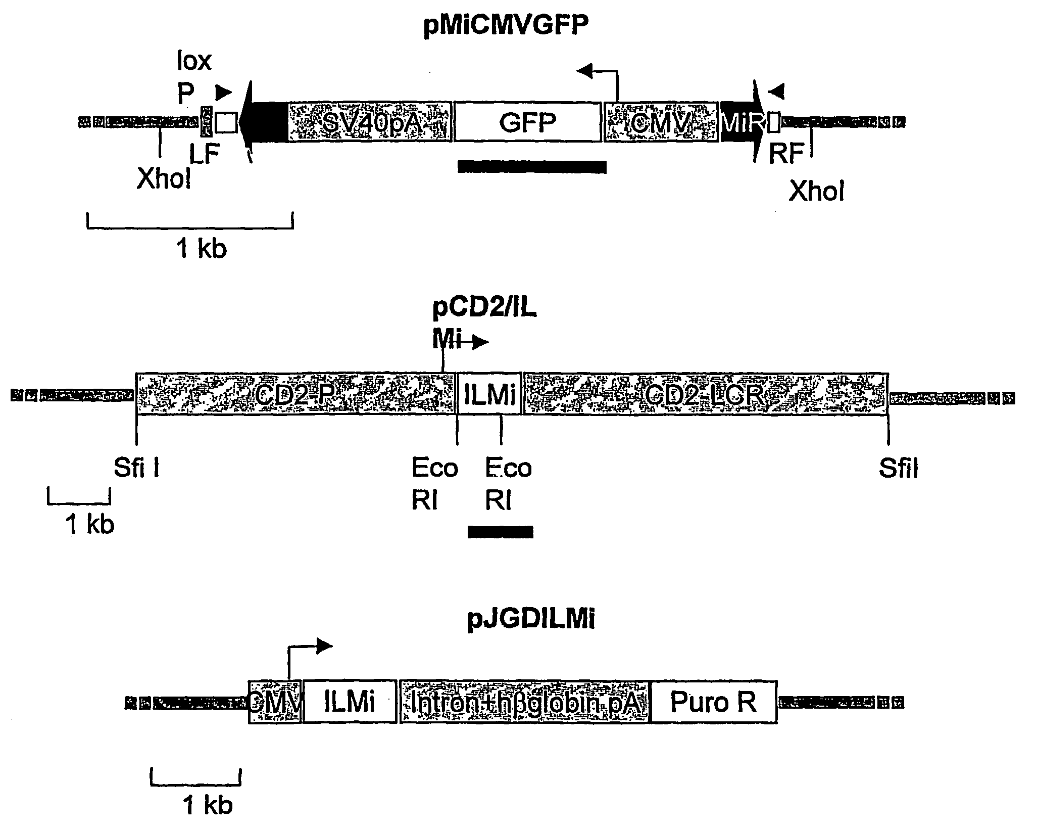
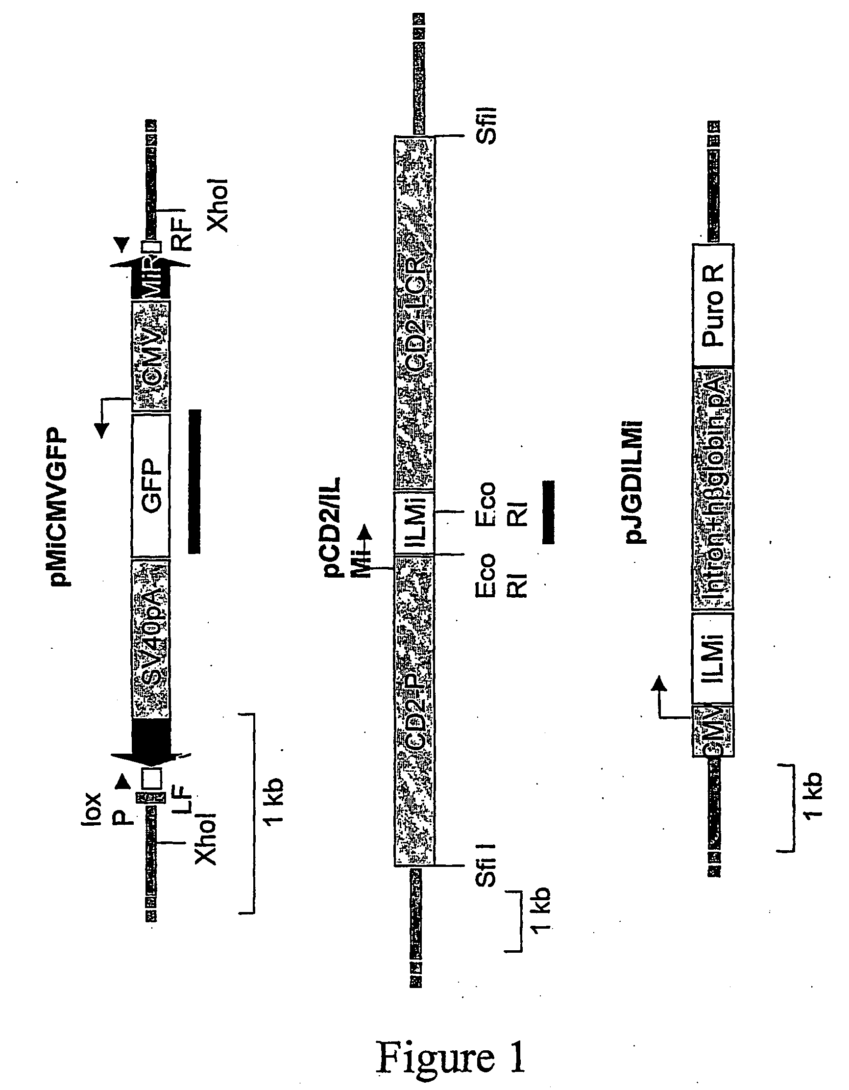
[0148]Two transgenic mouse lines are generated to determine whether Minos can transpose in mouse tissues: One containing a Minos transposon and another containing the Minos transposase gene expressed in a tissue-specific manner. The transposon-carrying line (line MCG) contains a tandem array of a fragment containing a Minos transposon (MiCMVGFP, FIG. 1) containing the GFP gene under the control of the cytomegalovirus promoter. The transposon is engineered such that almost all sequence internal to the inverted repeats is replaced by the CMV / GFP cassette. Not containing the transposase-encoding gene, this transposon is non-autonomous, and can only be mobilized when a source of transposase is present. The transposase-expressing line (line TM2) contains a tandem array of a construct comprising the Minos transposase cDNA under the control of the human CD2 locus, consisting of the CD2 promoter and LCR elements (pCD2 / ILMi, FIG. 1). In ...
example 2
Detection of Transposition in Cultured Embryonic Fibroblasts
[0151]The PCR excision assay is used to detect Minos excision in cultured embryonic fibroblasts carrying the MCG transgene. Cells are transfected with a plasmid carrying the Minos transposase cDNA under CMV control (pJGD / ILMi, FIG. 1) and analysed by the PCR excision assay. Excision products are detectable in transfected but not in non-transfected cells (data not shown). This result suggests that the transposon transgene is accessible to the Minos transposase in tissues other than T cells.
example 3
Detection of Excision Events
[0152]To determine the nature of the excision events, PCR products from thymus and spleen of MCG / +TM2 / +mice and from pJGD / ILMi transfected embryonic fibroblasts are cloned and sequenced. The sequence left behind after Minos excision in Drosophila consists of the TA dinucleotide duplication that is created upon Minos insertion, flanking the terminal 4 nucleotides of the transposon (i.e. either a AcgagT or a ActcgT insertion in the TA target site). In the mouse excisions analysed, the size and sequence of the footprints varies considerably (FIG. 4). Only 2 of the 32 footprints have the typical 6 bp sequence; the others contain extra nucleotides, in addition to complete or partial versions of the typical footprint. Four events have 1-2 nucleotides of the flanking D. hydei chromosomal sequence deleted. The differences in footprint structures observed between Drosophila and mouse may reflect the involvement of host factors in Minos excision and / or chromatid re...
PUM
| Property | Measurement | Unit |
|---|---|---|
| time | aaaaa | aaaaa |
| concentration | aaaaa | aaaaa |
| fluorescent | aaaaa | aaaaa |
Abstract
Description
Claims
Application Information
 - R&D
- Intellectual Property
- Life Sciences
- Materials
- Tech Scout
- Unparalleled Data Quality
- Higher Quality Content
- 60% Fewer Hallucinations
Browse by: Latest US Patents, China's latest patents, Technical Efficacy Thesaurus, Application Domain, Technology Topic, Popular Technical Reports.
© 2025 PatSnap. All rights reserved.Legal|Privacy policy|Modern Slavery Act Transparency Statement|Sitemap|About US| Contact US: help@patsnap.com


