Autoimmune disease biomarkers

a biomarker and autoimmune disease technology, applied in the field of autoimmune disease biomarkers, can solve problems such as unhealthy patients, and achieve the effect of improving anti-tnf therapies

Inactive Publication Date: 2008-10-16
INVITROGEN
View PDF5 Cites 58 Cited by
  • Summary
  • Abstract
  • Description
  • Claims
  • Application Information

AI Technical Summary

Benefits of technology

[0017]Another embodiment is a method of diagnosing RA in an individual comprising contacting a test sample from the individual with one or more target antigens and detecting binding of the one or more target antigens to one or more antibodies in the test sample, wherein the presence of the one or more antibodies against the one or more target antigens is indicative of rheumatoid arthritis, wherein the one or more target antigens are selected from the group comprising of Table 2 (as provided below) or a fragment thereof comprising an epitope.
[0018]Another embodiment is a method of diagnosing SLE in an individual comprising contacting a test sample from the individual with one or more biomarkers; and detecting binding of the one or more biomarkers to one or more antibodies in the test sample, wherein the presence of the one or more antibodies against the one or more biomarkers is indicative of SLE, wherein the one or more biomarkers are selected from the group comprising of Table 3 (as provided below) or a fragment thereof comprising an epitope.
[0019]Another embodiment is a method of diagnosing ANCA in an individual comprising contacting a test sample from the individual with one or more target antigens; and detecting binding of the one or more target antigens to one or more antibodies in the test sample, wherein the presence of the one or more antibodies against the one or more target antigens is indicative of ANCA, wherein the one or more target antigens are selected from the group comprising of Table 5 (as provided below) or a fragment thereof comprising an epitope.
[0020]Another embodiment of the present invention is a composition comprising one or more human antibodies from an individual with an autoimmune disease, wherein each antibody is bound to one or more target antigens each comprising an autoantigen of Table 1 or fragments thereof comprising an epitope. The target antigens may be immobilized on a solid support or may be part of a protein microarray. Another embodiment of the present invention is a solid support comprising two or more target antigens each comprising an autoantigen of Table 1 or fragments thereof comprising an epitope; and an immobilized human antibody control, wherein the human antibody control is a positive control for immunodetection.
[0021]The invention also provides kits that include one or more test antigens or one or more target antigens provided herein. The kits can include one or more reagents for detecting binding of an antibody from a sample. In some embodiments, the one or more test antigens or one or more target antigens of a kit are provided bound to a solid support. The invention includes kits that include biomarker detection panels of the invention, including biomarker detection panels in which the target antigens are bound to one or more solid supports. In some embodiments of kits, the kit provides a biomarker detection panel in which the target antigens of the detection panel are bound to a chip or array.
[0022]In some embodiments, the invention provides compositions, kits and methods for detecting one or more identified biomarkers as a diagnostic indicator for an autoimmune disease, such as RA, SLE, or ANCA. Additional uses of the invention include, among others: 1) the detection of one or more identified antigen biomarkers as a tool to select an appropriate therapeutic approach for treatment of a patient with a disease; 2) the use of one or more detected biomarkers as a vaccine candidate or therapeutic target; 3) the use of one or more identified biomarkers as a screening tool for use in the development of new therapeutics including antibodies; 4) the detection of one or more identified biomarkers as a tool for stratifying patients prior to infliximab (Remicade®) treatment; 5) the detection of one or more identified biomarkers for the early identification of the development of an SLE-like response in RA patients undergoing infliximab treatment; 6) the detection of observed anti-TNFα autoantibody response for the development of improved anti-TNF therapies; and 7) the detection of observed anti-TNFα autoantibody response as a surrogate marker for monitoring patient immune response to infliximab therapy.

Problems solved by technology

Numerous proteins on the array were bound by antibodies from patients diagnosed with RA, SLE and ANCA, but not healthy patients.

Method used


Image

  • Autoimmune disease biomarkers
  • Autoimmune disease biomarkers
  • Autoimmune disease biomarkers

Examples


example 1

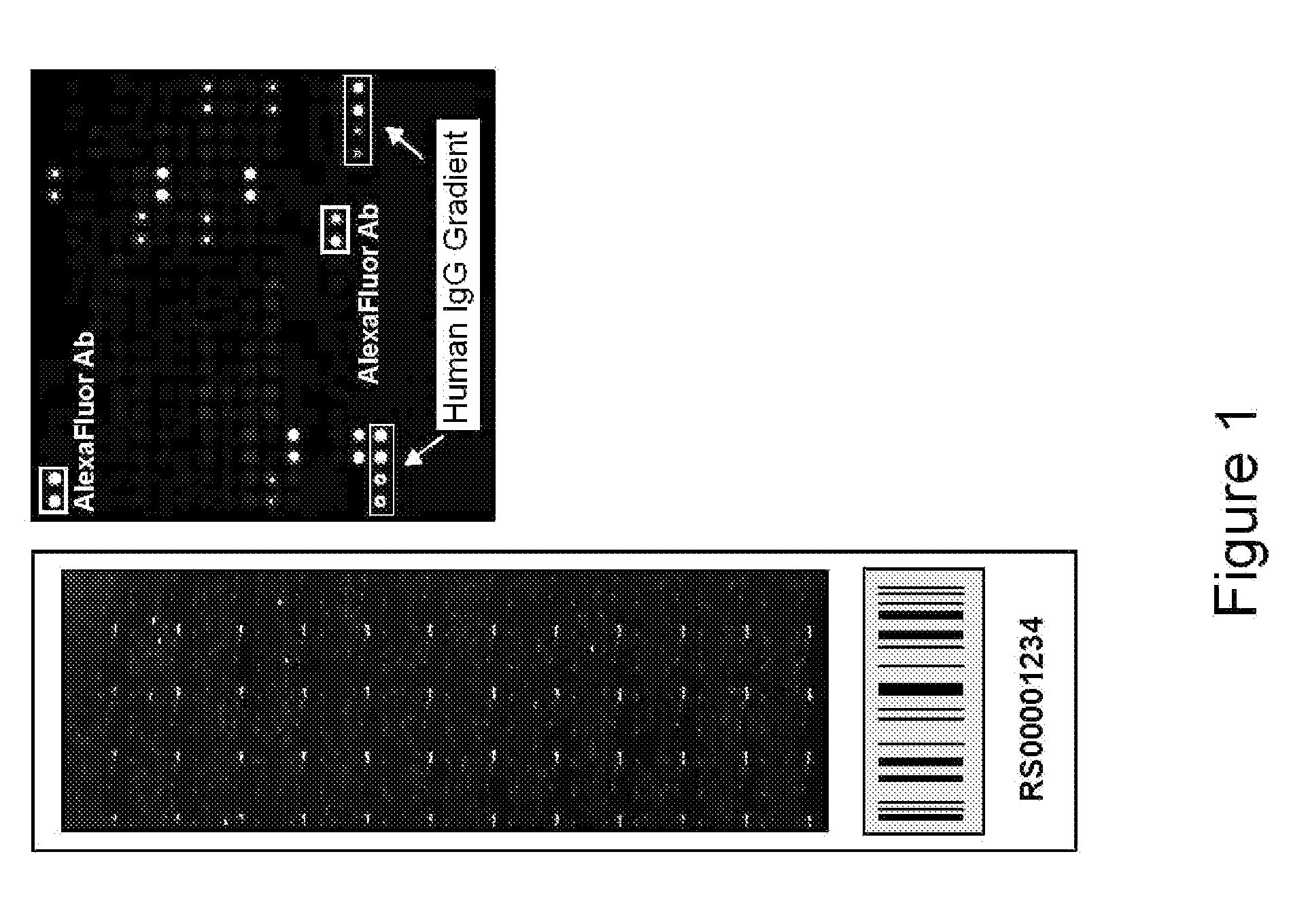
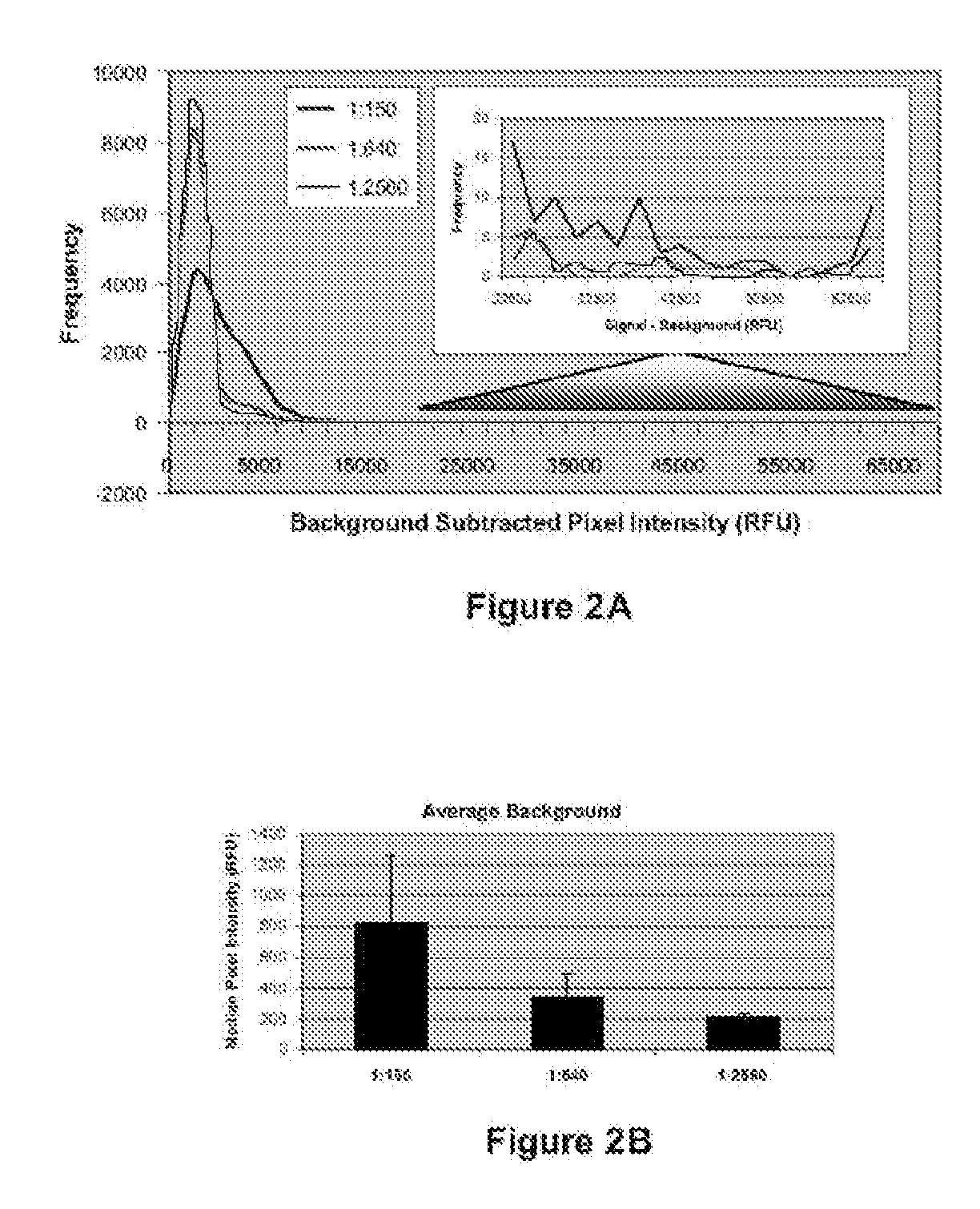
[0154]Serum from ten healthy control individuals, twelve individuals with RA prior to and following initiation of Remicade® treatment, twenty individuals with SLE, and twenty individuals with ANCA were profiled against a high throughput human protein array. Serum samples were diluted 1:150 and used to probe human ProtoArray™. Specifically, arrays were blocked for 1 hour, incubated with dilute serum solution for 90 minutes, washed 3×10 minutes, incubated with anti-human IgG antibody conjugated to AlexaFluor 647 for 90 minutes, washed as above, dried, and scanned. Following scanning, data was acquired using specialized software. Background-subtracted signals from each population were normalized utilizing a quantile normalization strategy. All possible pairwise comparisons were performed between all groups of samples included in the study utilizing an M-statistics algorithm in which the M-statistic is identified that is associated with the lowest possible p-value for a particular pairw...

example 2

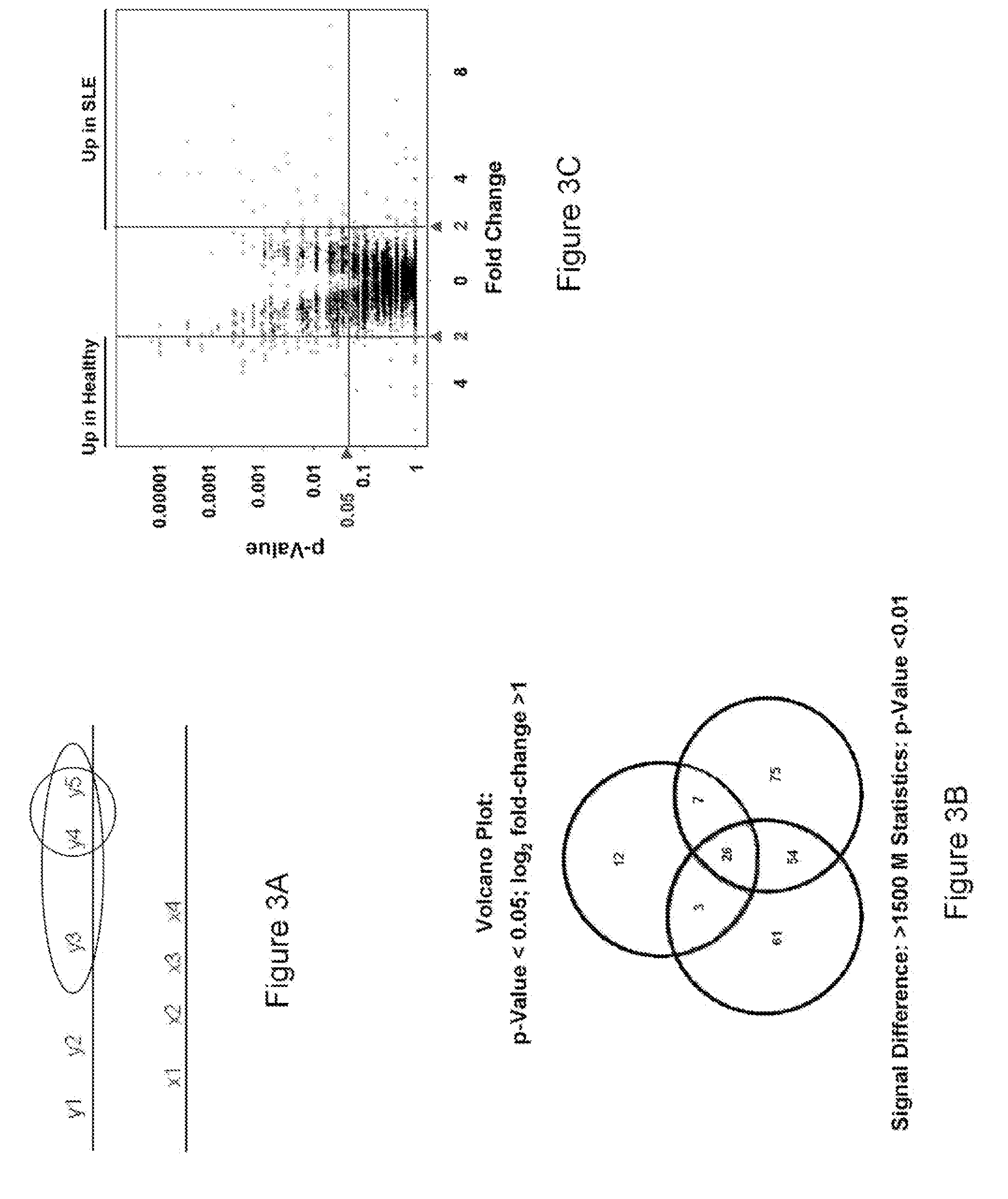
[0156]Serum samples from healthy individuals as well as individuals with autoimmune diseases including RA (Rheumatoid Arthritis), SLE (Systemic Lupus Erythrematosus) and ANCA (Anti-Neutrophil Cytoplasmic Antibody) were profiled on ProtoArray™ human protein microarrays as described in Example 1. Utilizing the calculations as described below, a number of potential antigen biomarkers were identified for autoimmune diseases. These proteins have the potential to serve as important diagnostic or prognostic indicators. Instead of an assay containing thousands or tens of thousands of proteins, a test sample can be profiled against an assay containing just the antigens associated with autoimmune disease, or a specific autoimmune disease. The tables below identify the autoantigens for RA, SLE, and ANCA.

[0157]Tables 1-7 identify antigens according to Genbank ID number for the nucleotide sequence that encodes the antigens. It is understood that an antigen of Tables 1-7 refers to a protein or fr...

example 3

[0164]Serum from twelve individuals with RA prior to and following initiation of infliximab (Remicade®) treatment were profiled against a high throughput human protein array as described in Example 1. Table 7A is a list of autoantigens that were bound by antibodies from RA patient sera and showed a decrease count after twenty weeks of infliximab treatment. Table 7B is a list of autoantigens that were bound by antibodies from RA patient sera and showed an increase count after twenty weeks of infliximab treatment.

TABLE 7ARA biomarkers showing a decrease count following treatment.Genbank IDnumber ofnucleic acidcoding for theproteinRA_T0RA_T20p-valueName or descriptionBC012105.1500.006192nuclear VCP-like, mRNABC025314.1600.001548immunoglobulin heavy constant gamma 1 (G1mmarker), mRNABC028039.1720.008454hypothetical protein MGC39900BC041037.1610.008978immunoglobulin heavy constant mu, mRNANM_003848.1500.006192succinate-CoA ligase, GDP-forming, beta subunit(SUCLG2), mRNANM_020367.2610.008...


PUM

PropertyMeasurementUnit
concentrationsaaaaaaaaaa
densityaaaaaaaaaa
timeaaaaaaaaaa

Abstract

Provided herein are novel panels of biomarkers for the diagnosis of autoimmune diseases, and methods and kits for detecting these biomarkers in samples of individuals suspected of having an autoimmune disease. Also provided are methods of monitoring the progression of an autoimmune disease and methods of monitoring the efficacy and side effects of a treatment for an autoimmune disease.

Description

CROSS-REFERENCE TO RELATED APPLICATIONS[0001]This application claims benefit of U.S. Provisional Application No. 60 / 867,022, filed Nov. 22, 2006, which is incorporated by reference in its entirety herein to the extent that there is no inconsistency with the present disclosure.BACKGROUND OF THE INVENTION[0002]This invention generally relates to biomarkers associated with autoimmune diseases, specifically Rheumatoid Arthritis (RA), Systemic Lupus Erythematosus (SLE) and Anti-Neutrophil Cytoplasmic Antibody (ANCA) associated diseases, and methods, compositions and kits for the diagnosis, prognosis, and monitoring the progression of autoimmune diseases.[0003]The development of autoantibodies is observed in autoimmune disorders and numerous cancers. Because of this, proteins targeted by autoantibodies (herein referred to as “autoantigens”) are effective biomarkers and form the basis of potential diagnostic and prognostic assays, as well as approaches for monitoring disease progression an...

Claims


Application Information

Patent Timeline
Patent Type & Authority Applications(United States)
IPC IPC(8): G01N33/564
CPCG01N33/564G01N2800/102G01N2800/104A61P19/02A61P29/00A61P37/06
Inventor MATTOON, DAWN R.SCHWEITZER, BARRYALCORTA, DAVIDPATEL, DHAVALKUMARFALK, RONALD
Owner INVITROGEN
Why Eureka
Try Eureka