
Hyperspectral fluorescence and absorption bioimaging

a bioimaging and hyperspectral fluorescence technology, applied in the field of chemical imaging of biological tissues and samples, can solve the problems of inability to inability to accurately detect the presence of spatial artifacts, so as to achieve the effect of increasing specificity

Inactive Publication Date: 2008-11-06
CHEMIMAGE
View PDF6 Cites 49 Cited by
  • Summary
  • Abstract
  • Description
  • Claims
  • Application Information

AI Technical Summary

Benefits of technology

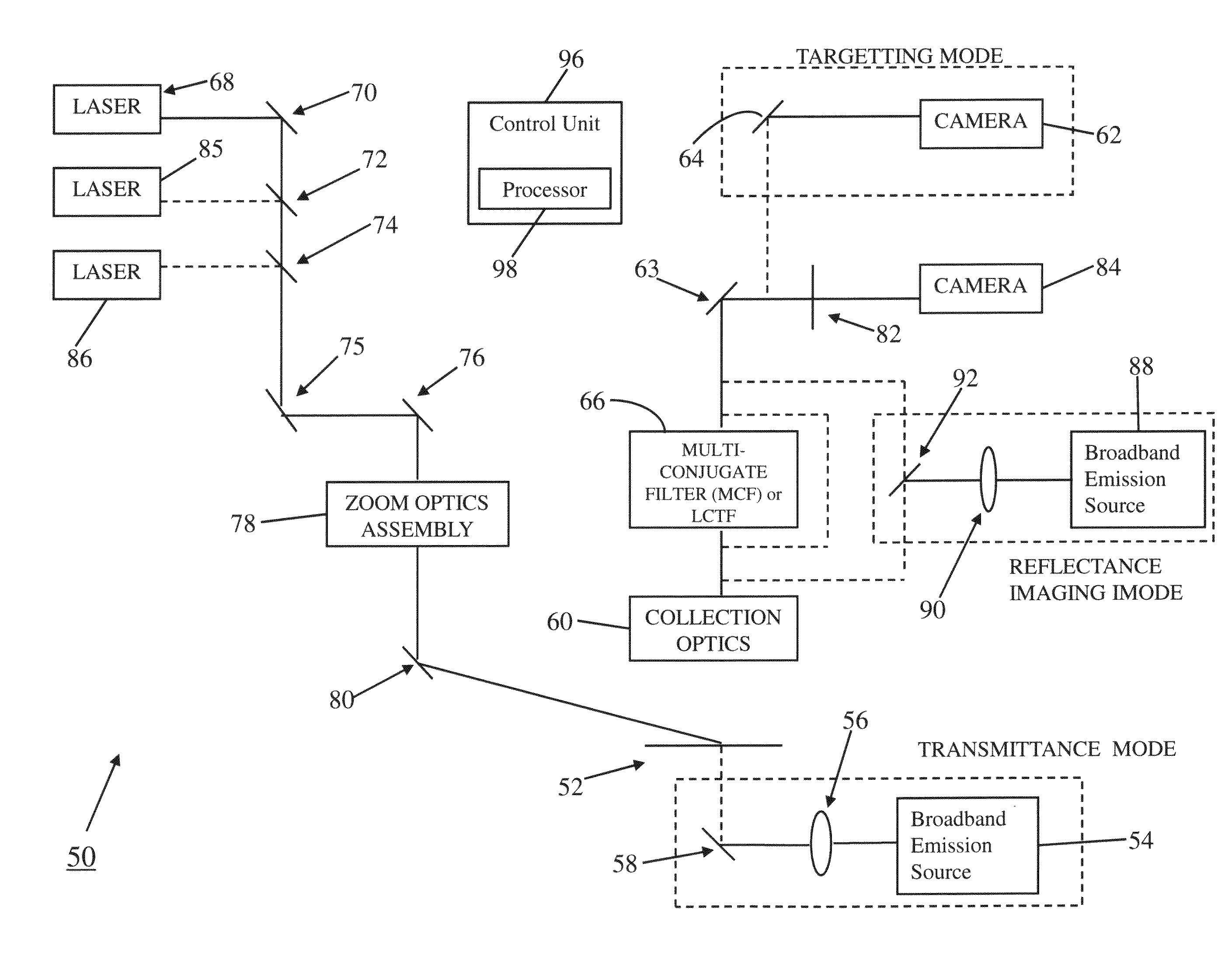
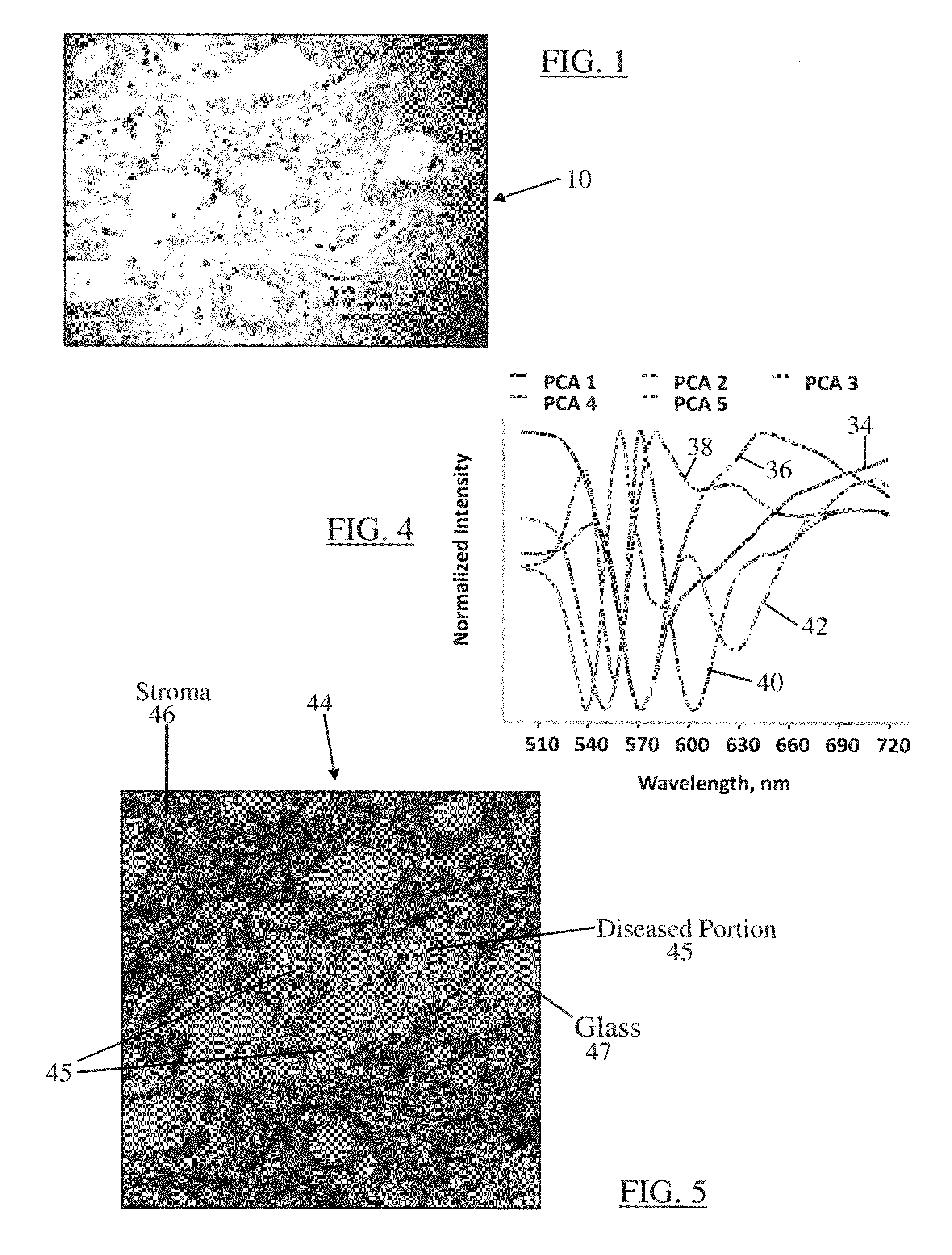
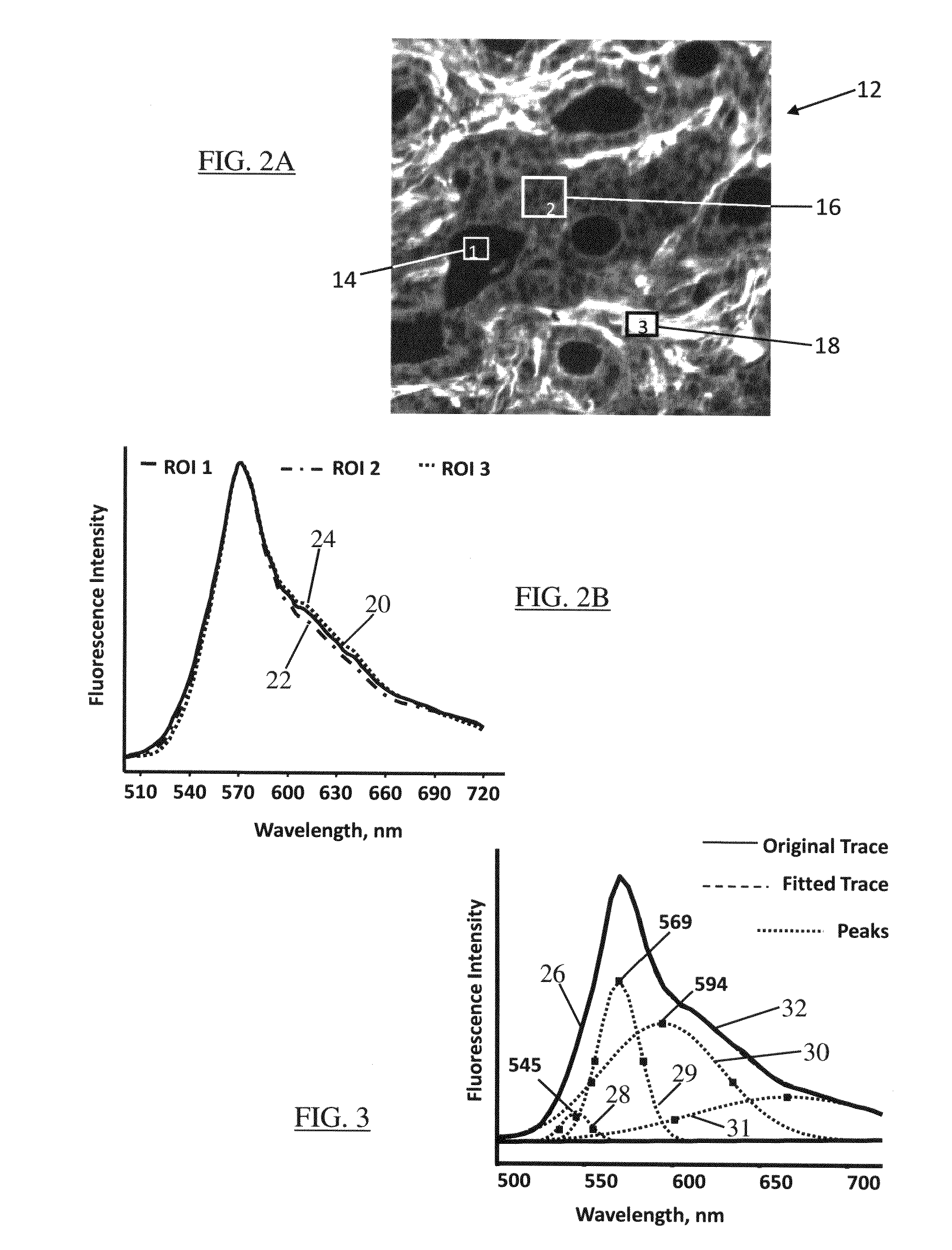
[0013]A system and method of hyperspectral chemical imaging (fluorescence or absorption based) according to one embodiment of the present disclosure provides an automated approach for a more detailed analysis of disease status of a biological sample. When a biological sample is labeled with a fluorescent or light-absorbing contrast-enhancing agent, interactions between the contrast-enhancing agent and one or more constituents (or cellular components) of the biological sample may be manifested through spectral contents of a plurality of regions in a hyperspectral chemical image of the sample. Observations of such manifestations through analysis of corresponding spectral contents may greatly assist a user (e.g., a pathologist) in detecting and differentiating diseased portions of the stained sample. Two-dimensional, wide-field chemical imaging may allow detection of multiple fluorescent or light-absorbing cellular probes (or cellular contaminants) with increased specificity, while accounting for non-uniform background fluorescence or absorption. Thus, hyperspectral chemical imaging may allow to identify multiple cellular components within the biological sample and to image their distribution within the sample, thereby assisting a pathologist to successfully and more accurately identify diseased portion(s) of the sample for further diagnosis and treatment.

Problems solved by technology

However, fluorescence microscopy suffers from certain known limitations such as, for example, presence of spatial artifacts, errors in quantitative measurements due to spectral bleed-through, reduction in detection sensitivity due to sample autofluorescence or reagent background fluorescence, effects of probe co-localization, or degradation in fluorescence image contrast.
However, such preliminary examination by a pathologist may not be sufficient or fully accurate in view of the limitations inherent in fluorescence microscopy.

Method used


Image

  • Hyperspectral fluorescence and absorption bioimaging
  • Hyperspectral fluorescence and absorption bioimaging
  • Hyperspectral fluorescence and absorption bioimaging

Examples


Embodiment Construction

[0023]The accompanying figures and the description that follows set forth the present disclosure in embodiments of the present disclosure. However, it is contemplated that persons generally familiar with optics, operation and maintenance of optical instruments (including spectroscopic instruments), or optical spectroscopy will be able to apply the teachings of the present disclosure in other contexts by modification of certain details. Accordingly, the figures and description are not to be taken as restrictive of the scope of the present disclosure, but are to be understood as broad and general teachings. In the discussion herein, when any numerical range of values is referred or suggested, such range is understood to include each and every member and / or fraction between the stated range of minimum and maximum. Furthermore, in the discussion below and in the accompanying figures, same reference numerals are used to describe same or similar elements, objects, or features.

[0024]The pr...


PUM


Abstract

A system and method of hyperspectral chemical imaging (fluorescence or absorption based) to provide an automated approach for a more detailed analysis of disease status of a biological sample. When a biological sample is labeled with a fluorescent or light-absorbing contrast-enhancing agent, interactions between the contrast-enhancing agent and one or more constituents (or cellular components) of the biological sample may be manifested through spectral contents of a plurality of regions in a hyperspectral chemical image of the sample. Observations of such manifestations through analysis of corresponding spectral contents may greatly assist a user (e.g., a pathologist) in detecting and differentiating diseased portions of the stained sample. Hyperspectral chemical imaging may allow to identify multiple cellular components within a biological sample and to image their distribution within the sample, thereby assisting a pathologist to successfully and more accurately identify diseased portion(s) of the sample for further diagnosis and treatment.

Description

REFERENCE TO RELATED APPLICATION[0001]The disclosure in the present application claims priority benefit under 35 U.S.C. § 119(e) of the U.S. Provisional Application No. 60 / 915,948, titled “Hyperspectral Fluorescence Bioimaging,” and filed on May 4, 2007, the disclosure of which is incorporated herein by reference in its entirety.BACKGROUND[0002]1. Field of the Disclosure[0003]The present disclosure generally relates to chemical imaging of biological tissues and samples and, more particularly, to a system and method to detect and differentiate diseased portions of a biological tissue or sample using hyperspectral fluorescence or absorption chemical imaging.[0004]2. Brief Description of Related Art[0005]Fluorescence is the result of a three-stage process that occurs in certain molecules called fluorophores or fluorescent dyes. In the first stage of the process, photon energy supplied from an external source such as an incandescent lamp or a laser diode is absorbed by the fluorophore, ...

Claims


Application Information

Patent Timeline
IPC IPC(8): G01J1/58
CPCG01J3/44G01N21/6452
Inventor TUSCHEL, DAVID
Owner CHEMIMAGE
Learn More