Eureka AIR delivers breakthrough ideas for toughest innovation challenges, trusted by R&D personnel around the world.

Microfluidic device and leucocyte antigen mediated microfluidic assay

a microfluidic and antigen-mediated technology, applied in fluorescence/phosphorescence, instruments, laboratory glassware, etc., can solve the problems of inability to detect leukocyte antigen, etc., to achieve the effect of convenient operation

Active Publication Date: 2009-01-08
ADVANCED ANIMAL DIAGNOSTICS
View PDF42 Cites 16 Cited by
  • Summary
  • Abstract
  • Description
  • Claims
  • Application Information

AI Technical Summary

Benefits of technology

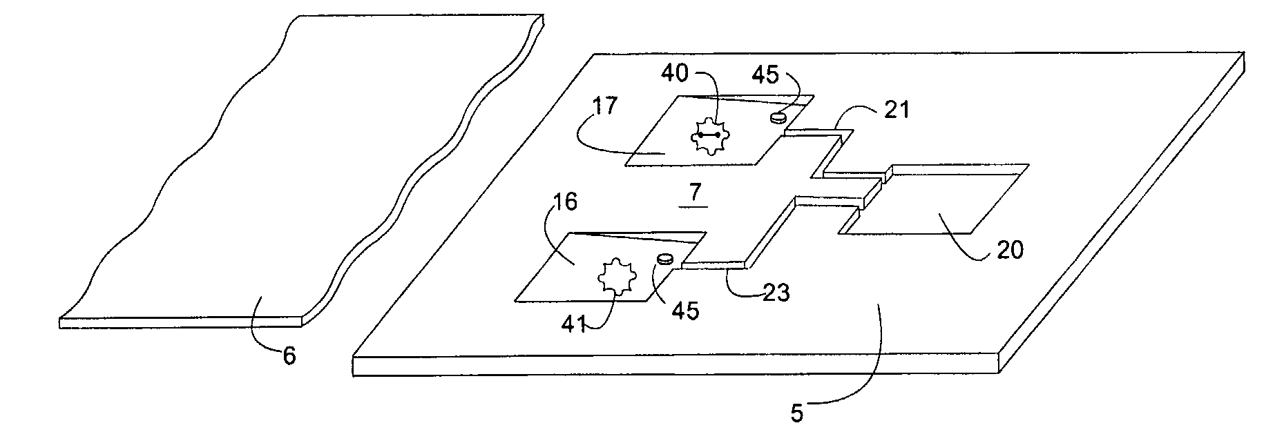
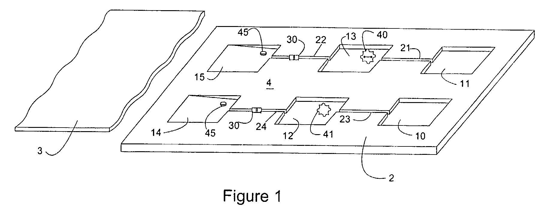
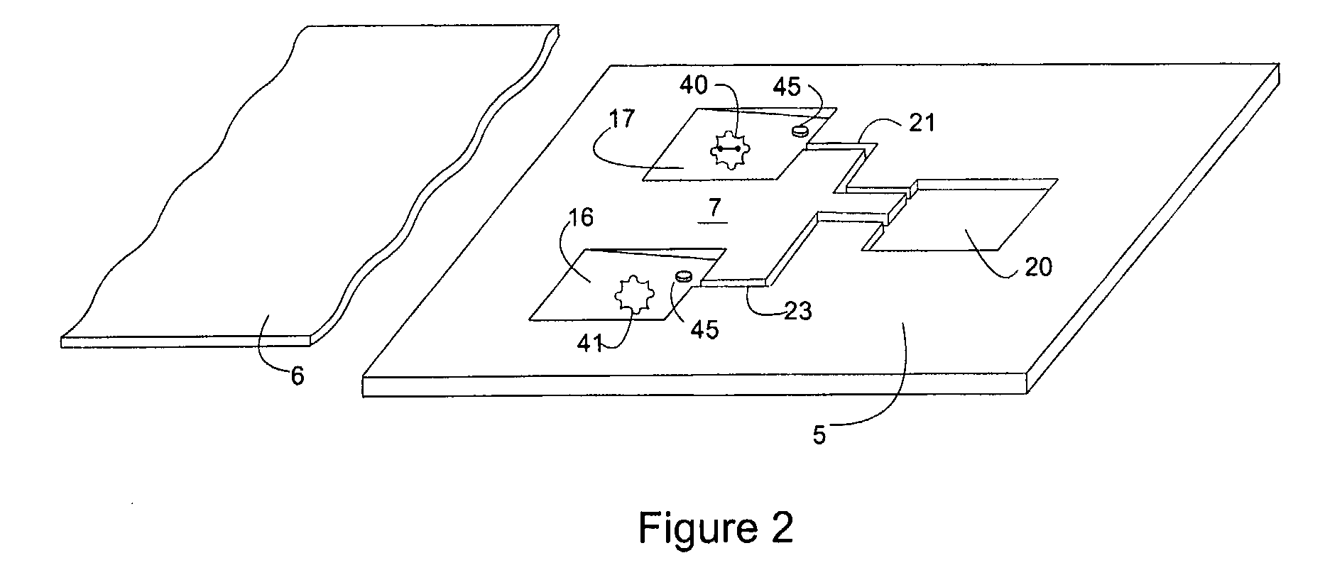
[0012]It is an object of the invention to provide both an assay and microfluidic device that can be used to test leucocyte containing body fluids such as undiluted whole blood for previous exposure to a predetermined antigen that is relatively quick, easy to do, accurate, wherein the device is disposable, the method is relatively safe, can be done in the lab or the field, uses very small volumes of blood, can test for multiple measurement factors or modes or tests for multiple antigens. The above objectives and more are achieved by the present invention and are distinct novel advantages as can be further seen from the disclosure herein.

Problems solved by technology

Serious problems exit however with antibody assays in this type of clinical testing.
Firstly, not all disease states, toxins or maladies produce a significant enough antibody response from B-lymphocytes to be useful in antibody assays.
A second problem with antibody tests arises when the time for production of antibodies, regardless of the quantity of antibodies produced, is sufficiently long such that by the time a diagnosis is made the patient may have suffered irreparable harm.
In antigen exposure situations where a subjects' medical treatment relies on a quick diagnosis, such low volume or long development time assays are essentially useless.
Besides these problems, antibody assays often require highly skilled technicians to perform them and they may need to be performed in a laboratory setting.
Not only does that mean the tests are costly, but it means that testing cannot be done on the site of an incident such as a terrorist attack, in the field or a doctors office, since samples must be sent back to the lab.
These types of tests also have several difficulties.
Further, the majority of the tests methods do not have the ability to accommodate more than a single mode i.e. a single measurement factor, of analysis.
A further problem has been that histochemical staining and cellular morphology are not capable of being done together with most methods.
Yet another problem with antibody assays are that T-lymphocytes represent a relatively small portion of leucocytes and even less a percentage of whole undiluted blood making observing or measuring them with small fluid samples extremely difficult.
The patent does not disclose comparing samples of the same cell types for differences in measurement factors, other than quantity of cells information it doesn't discuss any measurement factor observations at all and does not disclose use of this technique to identify cells which have been exposed to an antigen.
The patent further does not describe control methods and does not describe use in diagnosis of disease states other than inferring it can perform cell counts.

Method used


Image

  • Microfluidic device and leucocyte antigen mediated microfluidic assay
  • Microfluidic device and leucocyte antigen mediated microfluidic assay
  • Microfluidic device and leucocyte antigen mediated microfluidic assay

Examples


example

[0059]Latent Johne's Disease infection in dairy cattle. Johnes is the second costliest disease in the dairy industry and it is caused by the Mycobacterium avium paratuberculosis organism (MAP). The organism is known to be one of the slowest growers in the tuberculosis family, is extremely difficult to detect in latent asymptomatic infections, and it generates practically no antibody response in dairy cattle for months, or even years. However, upon initial exposure to cattle, MAP triggers an initial response in a subpopulation of bovine leukocytes (memory T-cells). The record of that initial encounter is retained in the immunological memory of the T Cell.

[0060]A bovine whole blood sample that been previously exposed to MAP is placed in the sample microchamber of the microfluidic device of the invention. The blood is then moved to the first reaction microchamber and re-stimulated using protein antigens that are unique to MAP, in combination with the antigen accelerator α2-macroglobuli...


PUM

PropertyMeasurementUnit
diameteraaaaaaaaaa
sizeaaaaaaaaaa
thicknessaaaaaaaaaa

Abstract

The present invention relates to an leucocyte antigen mediated microfluidic assay and a microfluidic device for analyzing a subjects' body fluids containing leucocytes to determine if the subject has been previously exposed to a predetermined antigen.

Description

[0001]This application claims priority of provisional application Ser. No. 60 / 570,543 filed May 13, 2004.BACKGROUND OF THE INVENTION[0002]1. Field of the Invention[0003]The present invention relates to an assay method and a microfluidic device for analyzing a subjects' body fluids containing leucocytes to determine if the subject has been previously exposed to the antigen. Specifically, the invention relates to an assay for observation, qualification and quantification of leucocyte measurement factors in a leucocyte containing fluid sample from a subject by exposure to a predetermined antigen in a novel microfluidic device in order to determine if there has been a prior exposure to the antigen.[0004]2. Description of the Related Art[0005]In leucocyte containing animals such as bird, mammals and especially humans, it is often necessary or determine if they have been previously exposed to a predetermined antigen. Antigens can be disease pathogens, allergy agents, toxins, infectious ag...

Claims


Application Information

Patent Timeline
Patent Type & Authority Applications(United States)
IPC IPC(8): C12Q1/02G01N21/00G01N21/64
CPCB01L3/5027B01L2300/0816B01L2300/0867G01N2015/008G01N33/56972G01N2333/8107B01L3/50273B01L2400/0406C12M23/58C12M23/16G01N2015/016C12M23/28C12M29/00G01N33/5091
Inventor RODRIGUEZ, RODOLFO R.DARR, DOUGLAS J.
Owner ADVANCED ANIMAL DIAGNOSTICS
Why Eureka
Eureka Blog
Learn More