System and Method for Robust Segmentation of Tubular Structures in 2D and 3D Images

a tubular structure and segmentation technology, applied in image enhancement, image analysis, instruments, etc., can solve the problems of less accurate corresponding results, more difficult tasks, and difficult segments of blood vessels, so as to avoid leakage, facilitate incorporation of user input, and fast computation

Inactive Publication Date: 2009-02-12
SIEMENS MEDICAL SOLUTIONS USA INC
View PDF3 Cites 13 Cited by
  • Summary
  • Abstract
  • Description
  • Claims
  • Application Information

AI Technical Summary

Benefits of technology

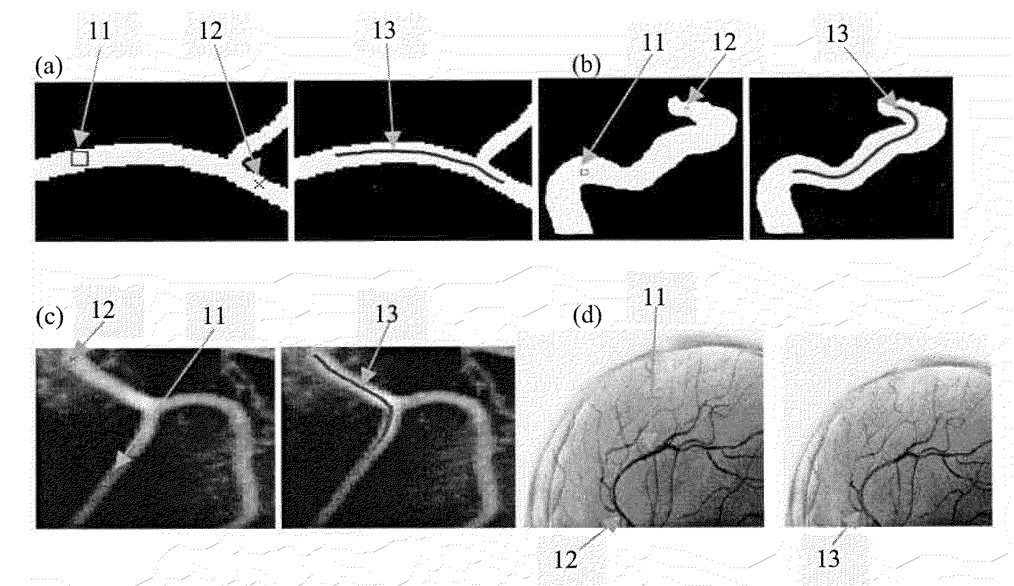
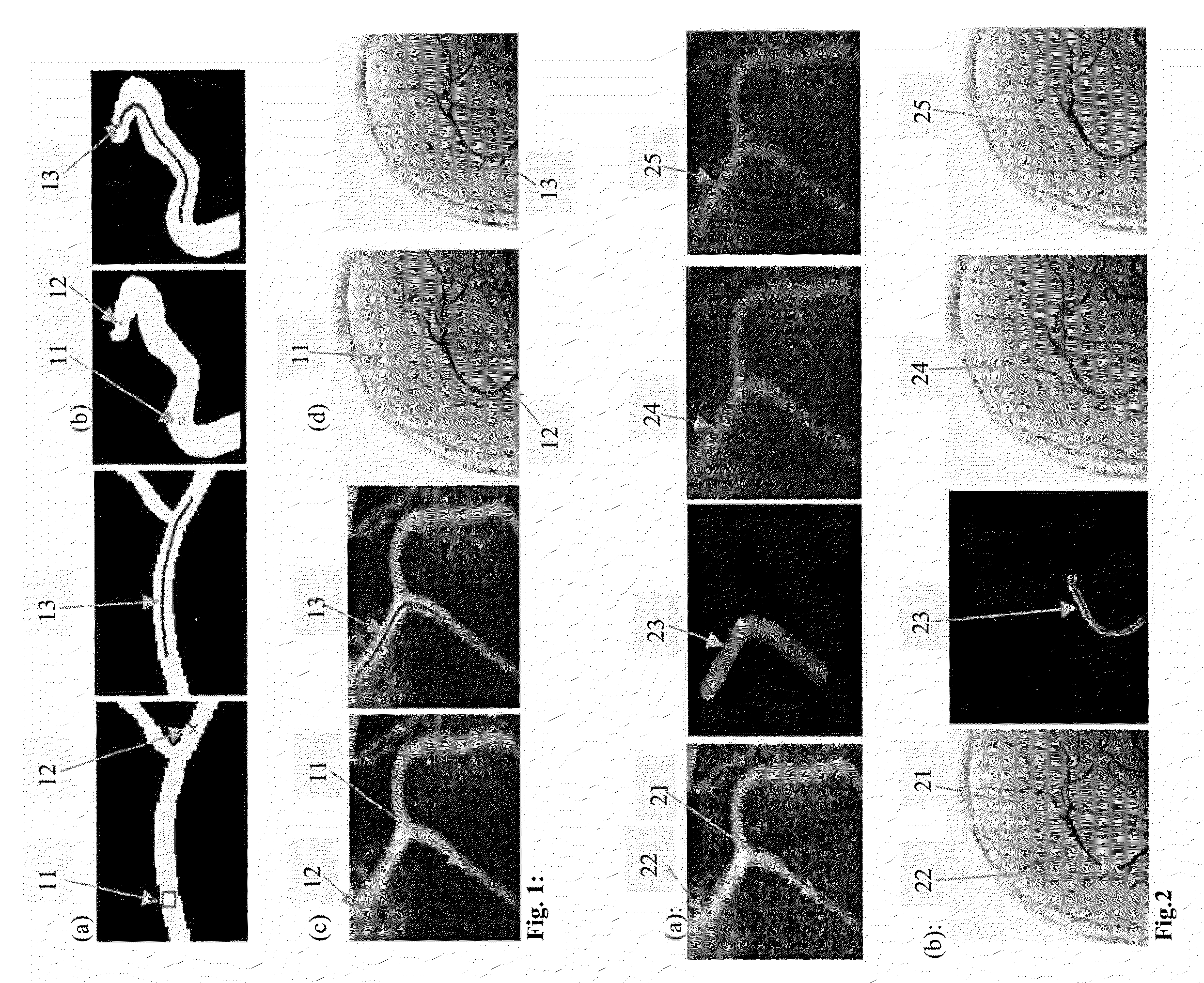
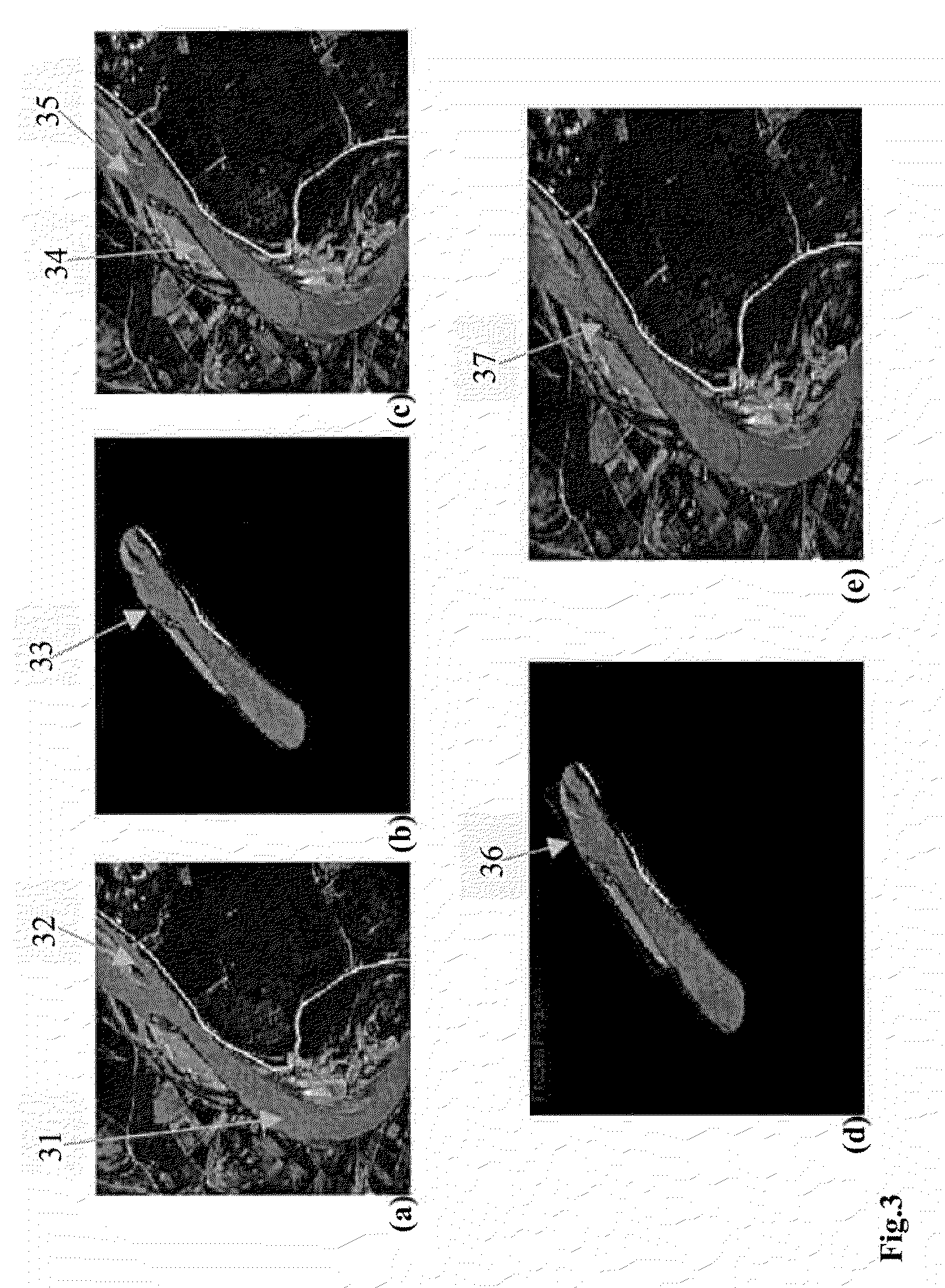
[0007]Exemplary embodiments of the invention as described herein generally include methods and systems for robust, interactive, semi-automatic extraction of the surface between two or more user-supplied end points for tubular- or vessel-like structures. A method according to an embodiment of the invention exploits features of minimal path techniques, such as global minima, fast computation, and ease of incorporating user input, to extract the shortest path between the user supplied end-points. This path is the global minimizer of an active contour model's energy along all possible paths joining the end-points. The extracted path is subsequently used to segment the vessel surface along the path. Some or all of the points forming the extracted path are used to automatically initialize a level set-based segmentation technique. This technique is only applied on the image portion consisting of the voxels that lie within a radius R from the extracted path. This step is rendered very fast because only a portion of the image along the path and within a radius R is considered. The width constraint also helps in avoiding any leakages that usually form at the root of the vessel, at bifurcations and branching areas, or close to the fast part of the evolving front. The radius R can be set by the user to a value that is proportional to the maximum width of the vessel segment along the path. To cope with any further leaks that may occur in the case of large variations of the vessel width between the user-supplied end-points, the points located at the neck of the potential leak areas are frozen during the course of evolution of the level set function. The points to be frozen can be determined from a few clicks by the user around the leak region. The potential of an algorithm according to an embodiment of the invention is demonstrated on several 2D / 3D synthetic and medical images. A typical application of a method according to an embodiment of the invention is the segmentation of vessels in medical images.

Problems solved by technology

Segmentation of blood vessels is a challenging task due to poor contrast, noise, as well as to their branching and changing geometry.
The task is even more challenging when a vessel surface along a particular path through branching vasculature is of interest.
The corresponding results may be less accurate due to the sensitivity of local gradients to noise and the chosen threshold.
However, the filter response may lead to incorrect results in case of a noisy intensity map.
These techniques, however, are sensitive to the initial placement of the vessel surface.
A bad initialization may result in several leakages out of the vessel boundaries.

Method used


Image

  • System and Method for Robust Segmentation of Tubular Structures in 2D and 3D Images
  • System and Method for Robust Segmentation of Tubular Structures in 2D and 3D Images
  • System and Method for Robust Segmentation of Tubular Structures in 2D and 3D Images

Examples


Embodiment Construction

[0026]Exemplary embodiments of the invention as described herein generally include systems and methods for segmenting tubular structures in digital medical images. Accordingly, while the invention is susceptible to various modifications and alternative forms, specific embodiments thereof are shown by way of example in the drawings and will herein be described in detail. It should be understood, however, that there is no intent to limit the invention to the particular forms disclosed, but on the contrary, the invention is to cover all modifications, equivalents, and alternatives falling within the spirit and scope of the invention.

[0027]As used herein, the term “image” refers to multi-dimensional data composed of discrete image elements (e.g., pixels for 2-D images and voxels for 3-D images). The image may be, for example, a medical image of a subject collected by computer tomography, magnetic resonance imaging, ultrasound, or any other medical imaging system known to one of skill in...


PUM


Abstract

A method for segmenting tubular structures in medical images includes providing at least a start point and an end point in a digital image volume, minimizing an action surface U0(p) which, at each image point p, corresponds to a minimal energy integrated along a path that starts at start point p0 and ends at p, sliding back on the minimal action surface from an end point to the start point to find a minimal path connecting the terminal points, initializing a level set function with points on the minimal path, and evolving the level set function to find a surface of a structure about the minimal path, wherein the level set function is constrained to be close to a signed distance function and wherein the level set function is prevented from growing wider than a predetermined diameter R, wherein the surface about the minimal path defines a tubular structure.

Description

CROSS REFERENCE TO RELATED UNITED STATES APPLICATIONS [0001]This application claims priority from “ROBUST SEGMENTATION OF TUBULAR STRUCTURES IN 2D AND 3D IMAGES”, U.S. Provisional Application No. 60 / 954,410 of Fahmi, et al., filed Aug. 7, 2007, the contents of which are herein incorporated by reference in their entireties.TECHNICAL FIELD [0002]This disclosure is directed to the segmentation of tubular structures in digital medical images.DISCUSSION OF THE RELATED ART [0003]The segmentation of vascular objects, such as blood vessels, human pulmonary tree, cerebral vasculature, aorta, etc., is important for various clinical tasks including surgical planning and diagnosis. Segmentation of blood vessels is a challenging task due to poor contrast, noise, as well as to their branching and changing geometry. The task is even more challenging when a vessel surface along a particular path through branching vasculature is of interest. Various approaches have been proposed for vessel segmentat...

Claims


Application Information

Patent Timeline
Patent Type & Authority Applications(United States)
IPC IPC(8): G06K9/00G06K9/34G06V10/26
CPCG06K9/34G06K9/6207G06T2207/30101G06T7/0089G06T2207/20116G06T7/0012G06T7/149G06V10/26G06V10/755
Inventor FAHMI, RACHIDWOLF, MATTHIASJEREBKO, ANNA
Owner SIEMENS MEDICAL SOLUTIONS USA INC
Why Eureka
Try Eureka