Apparatus And Method For Surgical Enhancement Of Aqueous Humor Drainage

a technology of aqueous humor and enhancement, applied in the field of apparatus and method for surgical enhancement of aqueous humor drainage, can solve the problems of blebs presenting a high incidence of post-surgical complications, surgical treatment, and progressive nerve damage and vision loss

Inactive Publication Date: 2009-02-12
ISCI INTERVENTIONAL CORP
View PDF100 Cites 233 Cited by
  • Summary
  • Abstract
  • Description
  • Claims
  • Application Information

AI Technical Summary

Problems solved by technology

Typically in glaucoma, the primary pathway for aqueous humor becomes narrowed or occluded, increasing IOP and resulting in gradual nerve damage and loss of vision.
Such conditions are usually treated by topical drugs in the form of eye drops, but may result in surgical treatment if drug treatment becomes ineffective or if patient compliance is an issue.
While blebs are effective in removing the aqueous humor, blebs present a high incidence of post-surgical complications due to irritation and infection.

Method used


Image

  • Apparatus And Method For Surgical Enhancement Of Aqueous Humor Drainage
  • Apparatus And Method For Surgical Enhancement Of Aqueous Humor Drainage
  • Apparatus And Method For Surgical Enhancement Of Aqueous Humor Drainage

Examples


example 1

[0050]An enucleated human cadaver eye was used for this test. The eye was prepared by removing any excess tissues around the limbus and replacing lost fluid until the eye was firm to the touch. A rectangular scleral flap, approximately 5 mm wide by 4 mm long, was incised posterior from the limbus in order to expose Schlemm's Canal.

[0051]A flexible microcannula prototype with an outside diameter of approximately 220 microns was used. The microcannula was fabricated with a communicating element comprised of polyimide tubing 0.006×0.008″ in diameter. Co-linear to the communicating element was a stiffening member comprised of a stainless steel wire 0.001″ diameter and a plastic optical fiber 0.004″ diameter. A heat shrink tubing of polyethylene terephthalate (PET) was used to bundle the elements into a single composite microcannula. The plastic optical fiber optic was incorporated to provide an illuminated beacon tip for localization. The beacon tip was illuminated using a battery power...

example 2

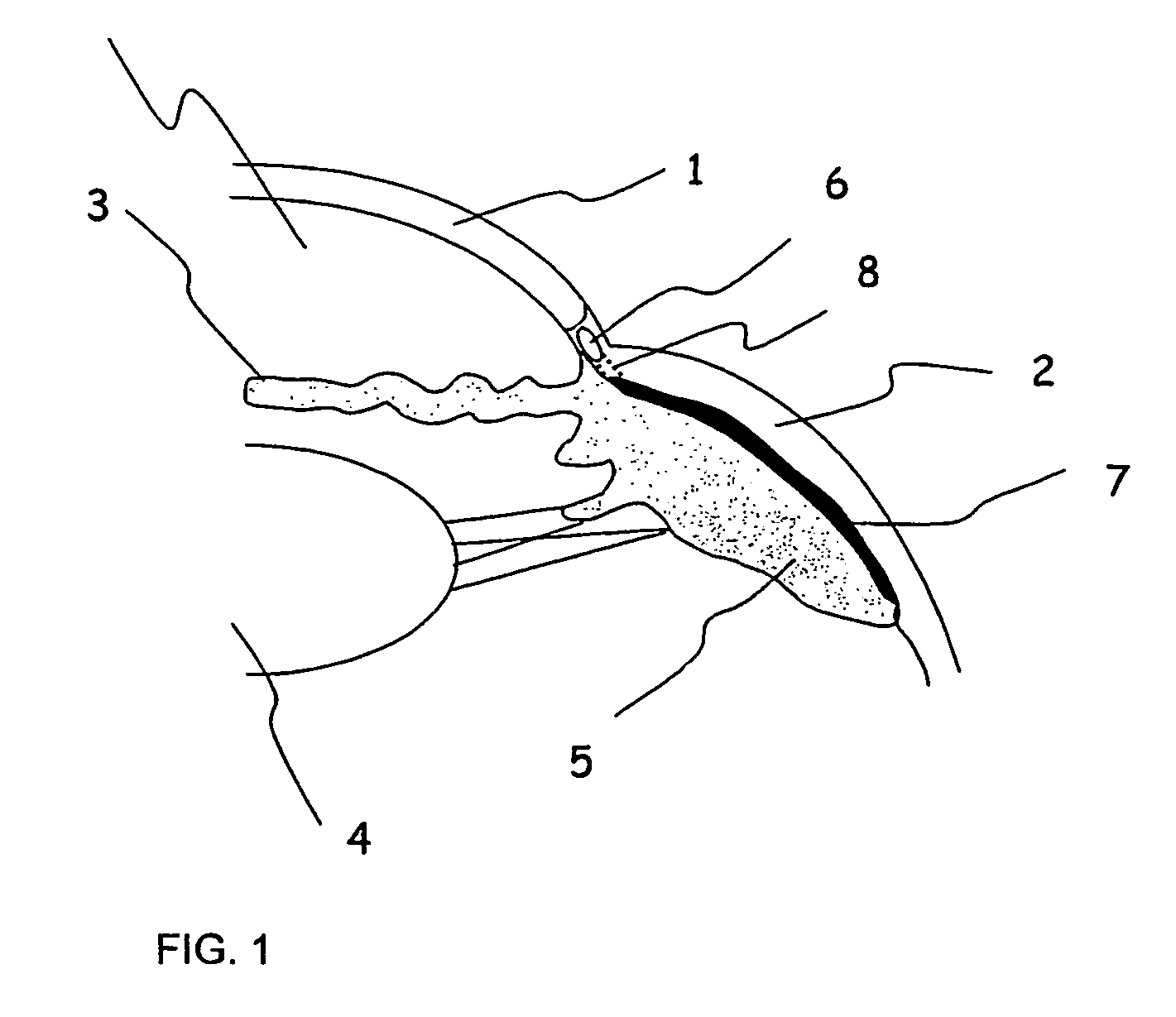
[0052]An enucleated human cadaver eye was prepared as in Example 1. Using a high resolution ultrasound imaging system developed by the applicant, an imaging scan was made of the tissues to plan the access and route for placing a shunt from the suprachoroidal space to the anterior chamber. A radial incision was made at the pars plana and extending through the sclera to expose the choroid. A microcannula (MicroFil, World Precision Instruments, Sarasota, Fla.) was employed. The microcannula comprised of a 34 gauge fused silica core tube coated with polyimide. The microcannula was inserted into the surgical incision in an anterior direction and at a small angle with respect to the scleral surface. The microcannula was advanced until the distal tip had penetrated into the anterior chamber. High resolution ultrasound imaging confirmed the cannula placement in the suprachoroidal space and extending above the ciliary body and penetrating the anterior chamber at the anterior angle. The dista...

example 3

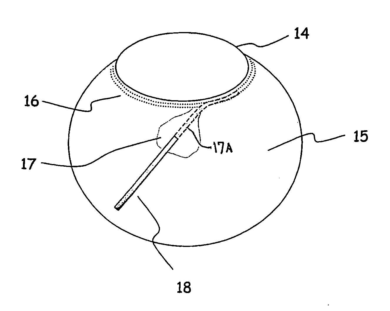
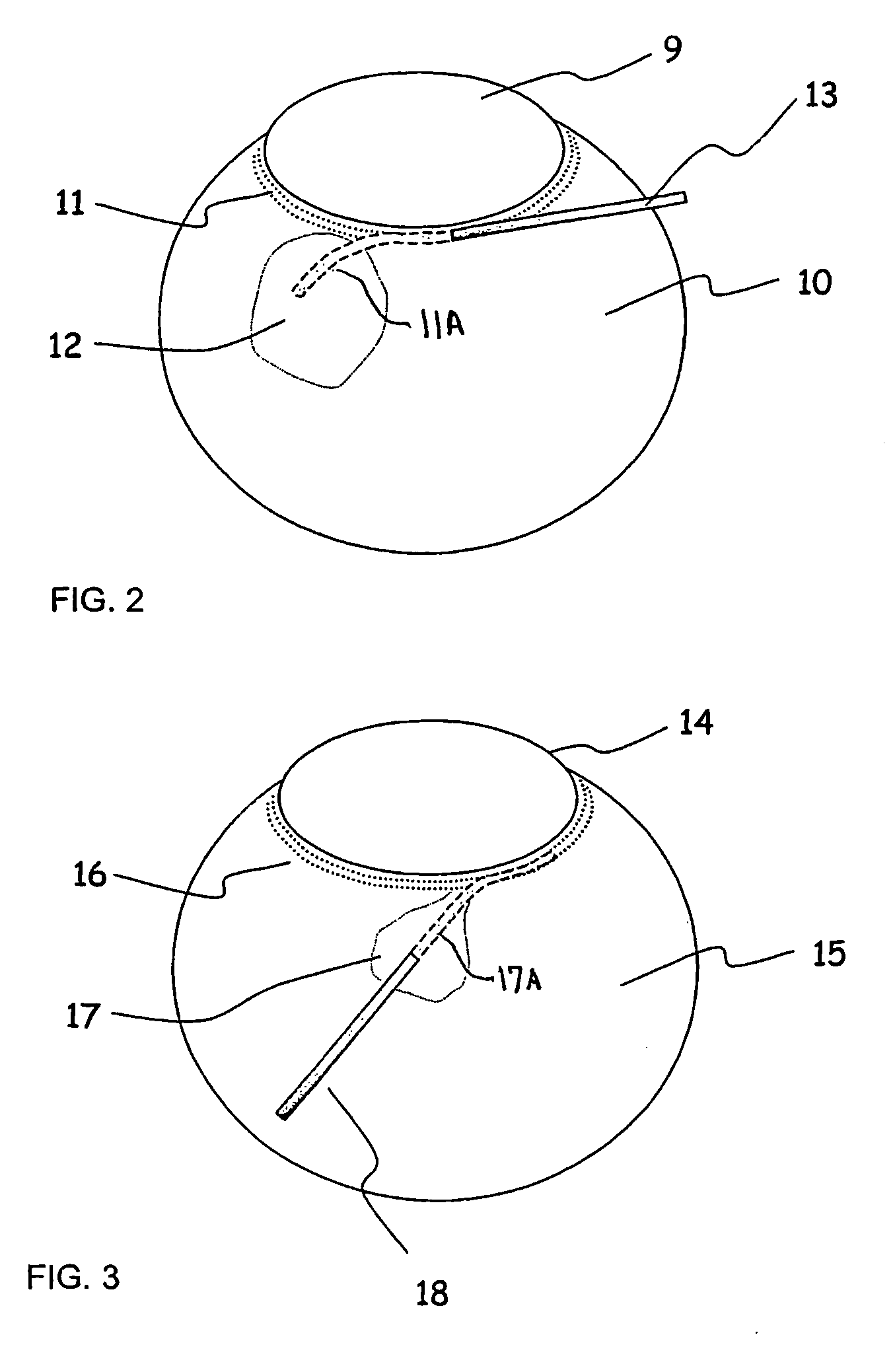
[0054]A test was performed to evaluate a tubular implant connecting Schlemm's Canal to the suprachoroidal space. An enucleated human cadaver eye was used and prepared as in Example 1. A radial incision was made in the superio-temporal limbal region, the incision extending for approximately 4 mm and to the depth of Schlemm's Canal. The posterior end of the incision was extended inward to expose the suprachoroidal without cutting into the choroid layer. The scleral spur fibers were left intact. A tubular implant comprised of polyimide tubing, 0.0044″ ID×0.0050″ OD was fabricated. The shunt tubing was split down the middle for a distance of 0.5 mm. The two halves of the tube were then folded back to create a “Tee” shaped flange at the distal end. A bend of approximately 30° was made in the body of the tube, 0.75 mm proximal to the split. The length of the base of the “Tee” was 2.5 mm. The proximal end of the shunt was placed firstly into the SCS, then the distal end was placed into the...


PUM


Abstract

An apparatus is provided for forming a tissue tract (8, 11A, 17A) from within a first passageway of an eye (11, 17) connecting to a second passageway in the eye (12, 16) comprising an elongated tool with a proximal end and distal end. The tool has an outer diameter in the range of about 50 to about 1000 microns. Methods of using the tool are provided for creating a fluid path for aqueous humor of an eye from a first passageway of the eye, such as the Schlemm's Canal, to a second passageway, such as the suprachoroidal space

Description

PRIORITY FROM RELATED APPLICATION[0001]Priority is hereby claimed from U.S. Provisional Application Ser. No. 60 / 567,024, filed Apr. 29, 2004, which is incorporated by reference in its entirety.BACKGROUND OF THE INVENTION[0002]Glaucoma is a disease condition of the eye in which increased intraocular pressure (IOP) is created by dysfunction in the drainage mechanism for the aqueous humor. Aqueous humor is produced within the eye in the ciliary body and flows within the anterior region of the eye. The aqueous humor normally drains primarily through a network of tissues at the interior angle of the anterior chamber, named the trabecular meshwork and subsequently into a circular drainage space named Schlemm's Canal. The aqueous humor continues its drainage path into collector channels and finally into aqueous veins to enter the venous system. This pathway for aqueous humor drainage is often called the trabeculo-canalicular pathway. The aqueous humor also drains through a more diffuse sec...

Claims


Application Information

Patent Timeline
Patent Type & Authority Applications(United States)
IPC IPC(8): A61M1/00A61F9/04A61B18/14A61B18/22A61F9/00A61F9/007A61M5/00
CPCA61B18/14A61F9/00781A61B18/22A61F9/00A61M5/00A61F9/008
Inventor CONSTON, STANLEY R.HEE, MICHAELKUPIECKI, DAVID J.MCKENZIE, JOHN R.YAMAMOTO, RONALD
Owner ISCI INTERVENTIONAL CORP
Why Eureka
Try Eureka