Method and apparatus for imaging target components in a biological sample using permanent magnets

a technology of permanent magnets and biological samples, applied in the direction of filtration separation, instruments, separation processes, etc., can solve the problems of low cost, inconsistent reliability, and inability to provide rapid and reliable methods

Active Publication Date: 2009-03-05
MENARINI SILICON BIOSYSTEMS SPA
View PDF9 Cites 37 Cited by
  • Summary
  • Abstract
  • Description
  • Claims
  • Application Information

AI Technical Summary

Problems solved by technology

Currently available methods do not provide a rapid, low cost and consistently reliable means for assessing a target population of cells by flow or image cytometry.

Method used


Image

  • Method and apparatus for imaging target components in a biological sample using permanent magnets
  • Method and apparatus for imaging target components in a biological sample using permanent magnets
  • Method and apparatus for imaging target components in a biological sample using permanent magnets

Examples


example 1

CD-Chex, Capture Efficiency

[0036]To determine the capture efficiency CD-Chex with known absolute numbers of leukocytes and their phenotypes is used.

Materials and Methods:

[0037]CD-Chex (lot #60650071):[0038]CD3+: 1859 / μl[0039]CD3+ / CD4+: 1221 / μl[0040]CD3+ / CD8+: 576 / μl

[0041]To 50 μl of CD-Chex, add 10 μl of CD3-FF (clone Cris7), 10 μl of CD4-APC and 10 μl of CD8-PE. After 25 minutes of incubation, 10 μl of this solution is injected into the chamber. PBS (1.8 ml) is added with 100 μl DAPI. The floater is then inserted. After capping, the chamber is placed on a rocker and rotated overnight (approximately 16 hrs). The chamber is inverted and the images of the floater are acquired.

Results:

[0042]For 100% capture efficiency, the floater surface contains:[0043]CD3+: 10328 cells[0044]CD3+ / CD4+: 6783 cells[0045]CD3+ / CD8+: 3200 cells

[0046]Images are acquired with different objectives and the resulting over-lay images are presented as shown in FIG. 6. FIG. 6A displays the image acquired using a 5...

example 2

Comparison Between Two Methods for Using Permanent Magnetics

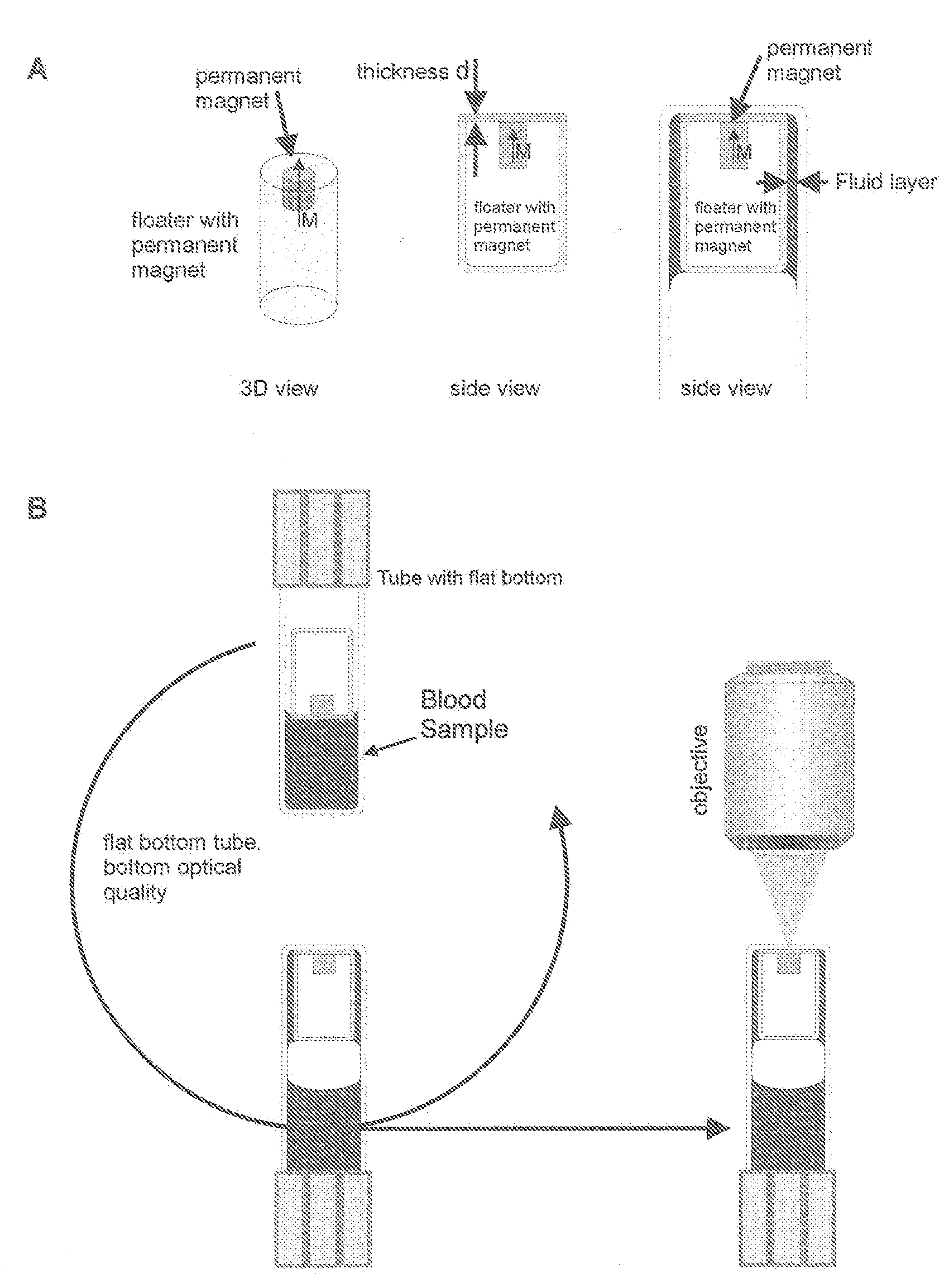
[0047]To determine the imaging of control cells after CellTracks® analysis using a method of inserting and removing a magnetic from the cell suspension (method 1) and using the method with a permanent magnet fixed to a floater (method 2) as described in the present invention.

Materials and Methods:

[0048]After analysis using the CellTracks® System, control cells from the CellTracks® cartridge were transferred to a chamber similar to FIG. 5B. The cartridge was washed several times with PBS using a pasteur pipette and all fluid used in the wash (approximately 500 microliters) was transferred to the chamber. Additional PBS was added to the volume to bring the total volume to 2 ml. Vial was placed on the tube rotator. The rotation speed was set so the floater moved through the entire fluid in one rotation. After complete mixing, 1.5 ml of control cells was injected into the vial together with 50 μl of Epcam ferrofluid and 10 μl o...

example 3

Amount of Ferrofluid

[0051]To determine the quality of image with increasing ferrofluid. As the amount of ferrofluid increases the image quality decreases. Cells become buried under a layer of ferrofluid and are invisible for detection. This results, in part, in the low recoveries.

Materials and Methods:

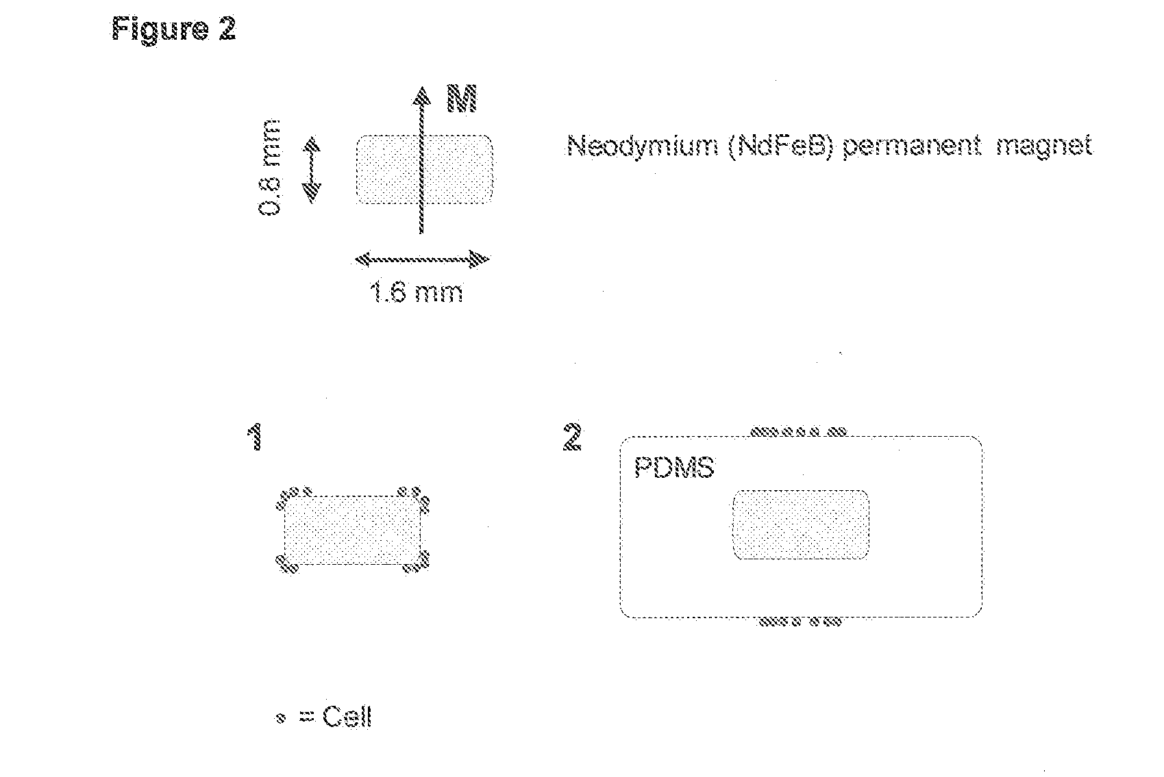
[0052]COMPEL Magnetic Microspheres, Dragon green, 2.914 107 / ml, diameter 8.44 microns, lot#6548 (Bangs Laboratories Inc. Catalog code UMC4F) were diluted 1:100. System buffer (1.5 ml) was added to the glass vial and 50 microliters containing 14570 beads were added together with 20, 40, 60 and 80 microliters of EpCam ferrofluid (20 mg / ml). Fluorescence images were acquired after 15 and 30 minutes of rotation. Test tube rotator was set at 10 rpm, resulting in 150 and 300 rotations.

[0053]Floater is Corning 1 / 16″ diameter magnet.

Results:

[0054]Images are acquired with a 5× and 40× objectives. As shown in FIG. 8, 5× and 40× objectives were used to image 20, 40, 60 and 80 microliters of EpCam...


PUM

PropertyMeasurementUnit
heightaaaaaaaaaa
diameteraaaaaaaaaa
thicknessaaaaaaaaaa

Abstract

A system for enumeration of cells in fluids by image cytometry is described for assessment of target populations such as leukocyte subsets in different bodily fluids or bacterial contamination in environmental samples, food products and bodily fluids. Briefly, fluorescently labeled target cells are linked to magnetic particles or beads. In one embodiment, a small, permanent magnet is inserted directly into the chamber containing the labeled cells. The magnets are coated with PDMS silicone rubber to provide a smooth and even surface which allows imaging on a single focal plane. The magnet is removed from the sample and illuminated with fluorescent light emitted by the target cells captured by a CCD camera. In another embodiment, a floater having a permanent magnet allows the target cells to line up along a single imaging plane within the sample solution. Image analysis can be performed with a novel algorithm to provide a count of the cells on the surface, reflecting the target cell concentration of the original sample.

Description

CROSS REFERENCE TO RELATED APPLICATIONS[0001]This application is a non-provisional application, which is incorporated by reference herein and claims priority, in part, of U.S. Provisional Application No. 60 / 594,198, filed Mar. 19, 2005, now abandoned, and is a continuation-in-part of U.S. Ser. No. 11 / 897,471, filed Aug. 30, 2007.FIELD OF THE INVENTION[0002]The invention relates generally to imaging target components in a fluidic (biological) sample. More specifically, methods and apparatus are described that provide for the positive selection of target cells in a blood sample. Small permanent magnets are added directly to a blood sample containing CD4 immunomagnetic labeled fluorescently stained Acridine Orange (AO) whole blood.BACKGROUND ART[0003]The use of immunomagnetic separation technology provides greater sensitivity and specificity in the detection of target entities in blood for example, but not limited to, intact circulating cancer cells and endothelial cells. This simple a...

Claims


Application Information

Patent Timeline
Patent Type & Authority Applications(United States)
IPC IPC(8): C12Q1/06C12M1/00
CPCG01N33/54326G01N33/56972G01N2446/00G01N2333/70514G01N33/582
Inventor TIBBE, ARJAN G.J.TERSTAPPEN, LEON W.M.M.
Owner MENARINI SILICON BIOSYSTEMS SPA
Why Eureka
Try Eureka