Optical wavelength range for high contrast imaging of cancer

a high contrast, optical technology, applied in the field of optical imaging of cancerous and precancerous tissue, can solve the problems of inability or inability to bring a probe into direct contact with the tissue needing to be imaged, prone to size limitations, and difficult or impossible in vivo detection of many cancer tissues by the transillumination apparatus, etc., to achieve the effect of higher or lower scattering coefficien

Inactive Publication Date: 2009-03-19
THE GENERAL HOSPITAL CORP
View PDF20 Cites 3 Cited by
  • Summary
  • Abstract
  • Description
  • Claims
  • Application Information

AI Technical Summary

Benefits of technology

[0019]In accordance with another aspect of the invention, the filtering step includes passing only light within a wavelength range in which cancerous tissue and healthy tissue have absorption coefficients that are approximately equal, and in which cancerous tissue has a higher or lower scattering coefficient than healthy tissue, causing cancerous tissue to appear darker or brighter than healthy tissue. It is also contemplated that the steps of illuminating and constructing an image can be performed in a confocal imaging section of a confocal microscope. The step of illuminating can include transmitting illumination light between the confocal imaging section and a remote probe that is insertable into a body. It is also possible for the step of illuminating to include polarizing the illumination light, and for the registering step to include registering two reflected polarization components to be processed into an image.

Problems solved by technology

However, the transillumination apparatus makes in vivo detection of many cancer tissues difficult or impossible because the illumination light must project from one side of the tissue, while the detection device (e.g., camera) must be on the opposite side of the tissue in order to receive the light from the illumination means through the tissue sample.
However, the apparatus requires catalogued data, which may or may not be available for a given tissue type or for cancer within the tissue type.
Moreover, it is not always possible or desirable to bring a probe into direct contact with tissue needing to be imaged.
However, since the apparatus measures transmitted and / or scattered light from a tissue sample, it is prone to size limitations similar to those in U.S. Pat. No. 5,451,785, described above, when imaging of in vivo tissue is required.
However, this technique requires use of a spectrometer, which adds bulk and cost to the system.
Therefore, normalization of the green fluorescence images using NIR reflectance is not accurate.
However, many of the known methods are difficult or impossible to apply to in vivo imaging of cancer tissue.

Method used


Image

  • Optical wavelength range for high contrast imaging of cancer
  • Optical wavelength range for high contrast imaging of cancer
  • Optical wavelength range for high contrast imaging of cancer

Examples


Embodiment Construction

[0040]Reference will now be made in detail to the present preferred embodiments of the invention, examples of which are illustrated in the accompanying drawings. The method and corresponding steps of the invention will be described in conjunction with the detailed description of the system. The devices and methods presented herein may be used for imaging and diagnosing cancer tissues. The present invention is particularly suited for in vivo optical imaging of cancerous tissues in which cancerous tissue is contrasted with adjacent healthy tissue.

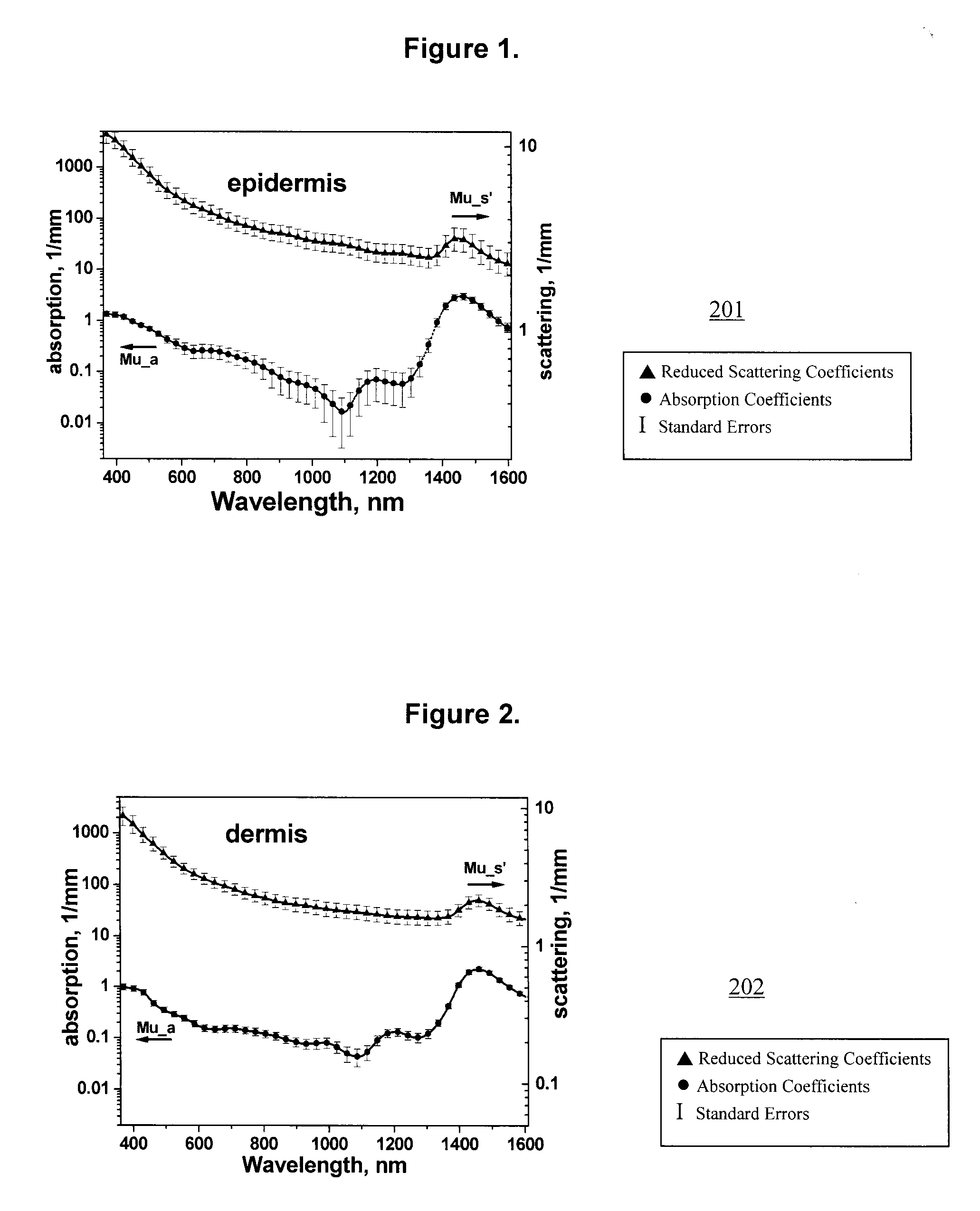
[0041]Differences in absorption and / or scattering of cancerous and normal skin have the potential to provide a basis for non-invasive cancer detection. In accordance with the invention, it was discovered that there are significant differences in the scattering of cancerous and healthy tissues in the spectral range from 1050-1400 nm. In this spectral region, the scattering dominates the absorption by at least one order of magnitude, the scatte...


PUM


Abstract

An apparatus for imaging cancer in which cancer tissue is contrasted against adjacent healthy tissue. The apparatus includes illumination and, possibly polarization means configured to provide illumination light on a region of tissue. Detection means are optically coupled to the illumination means, the detection means being adapted to detect reflected light from the tissue (and its polarization state, if applicable) when the tissue is illuminated by the illumination means. Filtering means are optically coupled to the detection means. The filtering means are configured to filter the illumination light and/or reflected light to pass only light within a range, or any subrange between about 1050 to about 1400 nm. Methods for imaging cancer contrast cancer tissue against adjacent healthy tissue.

Description

CROSS-REFERENCE TO RELATED APPLICATION[0001]This application claims priority to U.S. Provisional Patent Application No. 60 / 994,038 filed Sep. 17, 2007, which is incorporated herein by reference.BACKGROUND OF THE INVENTION[0002]1. Field of the Invention[0003]The present invention relates to optical imaging of cancerous and precancerous tissue. Particularly, the present invention is directed to optical imaging of dysplastic and cancer tissue within a wavelength range in which cancer tissue is contrasted with adjacent healthy tissue.[0004]2. Description of Related Art[0005]A variety of devices and methods are known in the art for detecting cancer tissue. Of such devices, many are directed to optical detection of cancer tissues. Nonmelanoma skin cancers including basal cell carcinomas (BCC) and squamous cell carcinomas (SCC), are more common than all other types of human cancers. These cancers have an intrinsically low optical contrast in the visible spectral range. A number of techniqu...

Claims


Application Information

Patent Timeline
Patent Type & Authority Applications(United States)
IPC IPC(8): A61B6/00
CPCA61B5/0068A61B5/446A61B5/444A61B5/06
Inventor YAROSLAVSKY, ANNA N.SALOMATINA, ELENA
Owner THE GENERAL HOSPITAL CORP
Why Eureka
Try Eureka