
Connective Tissue Closure Device and Method

a technology of connective tissue and closure device, which is applied in the field of medical devices, can solve the problems of affecting the patient's recovery, and affecting the healing effect of patients, and can take hours for a clot to form

Inactive Publication Date: 2009-04-02
BOSTON SCI SCIMED INC
View PDF37 Cites 24 Cited by
  • Summary
  • Abstract
  • Description
  • Claims
  • Application Information

AI Technical Summary

Benefits of technology

The patent describes a wound closure device that includes an expander and a fabric made from polylactic acid fiber. The device can be used to close wounds in blood vessels. The device is inserted through an introducer sheath and the expandable frame is expanded to secure the fabric around the blood vessel. The fabric has microhooks that engage with the adventitia surrounding the blood vessel. A cinch ring is then slid down around the fabric and a suture is connected to the fabric to close the wound. The device can be expanded and contracted using a deployment mechanism. The technical effects of this device include improved wound closure and reduced risk of complications.

Problems solved by technology

While effective, the process can be relatively slow, often taking up to 24 hours for a clot to be formed.
In addition, during this time, the patient must remain stationary In addition, the simple application of such pressure or weight is often of great discomfort to the patient, or can detrimentally affect other systems of the body due to the applied weight, pressure and immobility.
While this is also effective, such devices and methods do have the unfavorable disadvantage of positioning a portion of the closure within the blood vessel itself.
While these latter devices and methods can be effective, they are often difficult for the physician to execute, can cause pain to the patient, and can be relatively time consuming Accordingly, it would be advantageous to provide an apparatus or method for closing the opening in the blood vessel which is immediate and relatively easy for the physician to execute, but which does so without leaving any structure behind, or at least using structure which is bioabsorbable after a relatively short period of time, thereby ultimately leaving no foreign substance in the body

Method used


Image

  • Connective Tissue Closure Device and Method
  • Connective Tissue Closure Device and Method
  • Connective Tissue Closure Device and Method

Examples


Embodiment Construction

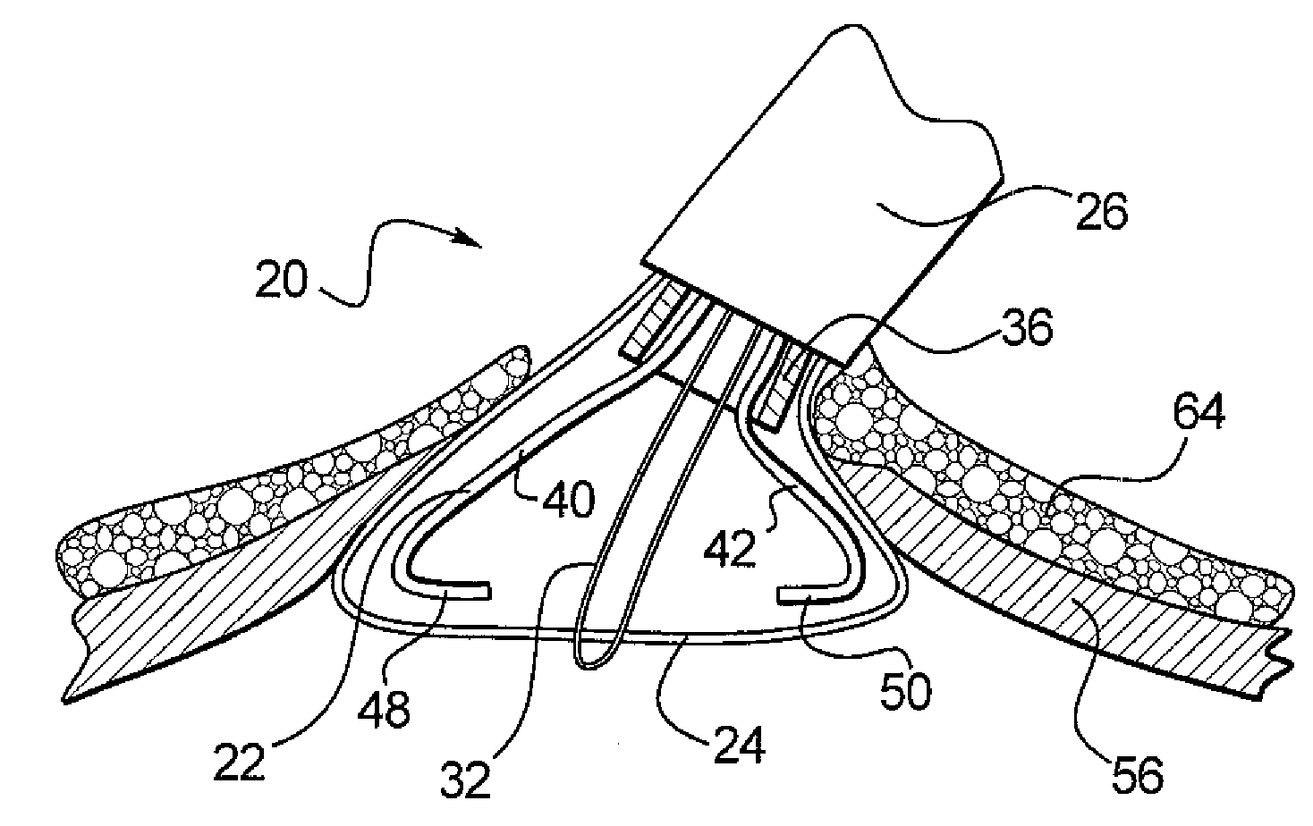
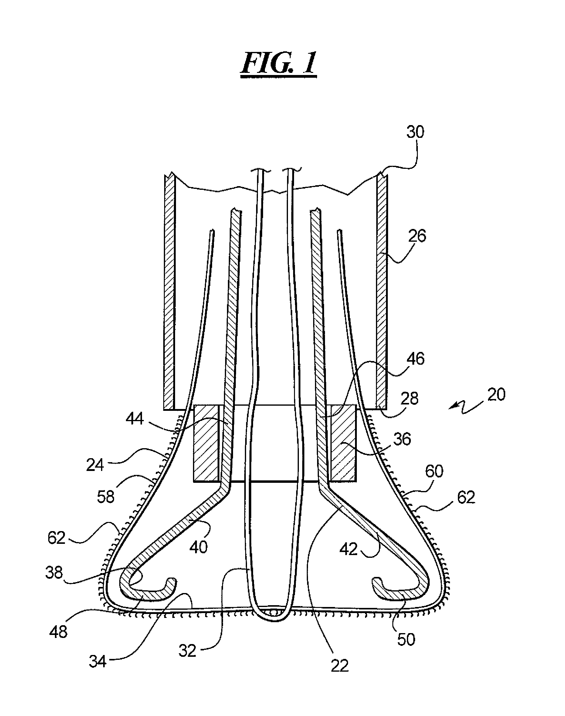
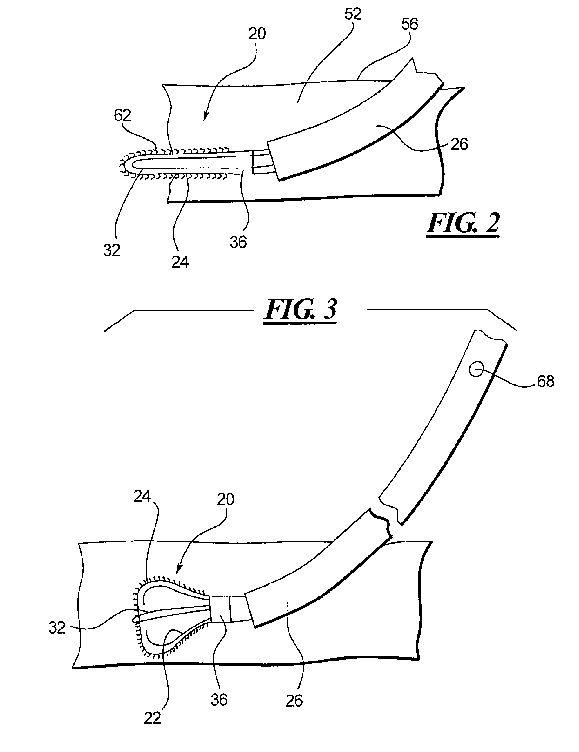
[0025]Referring to the drawings and with specific reference to FIG. 1, a wound closure device constructed in accordance with the teachings of the disclosure is generally referred to as reference numeral 20. It is to be understood that while the device 20 is described below in connection with the closure of an opening in a blood vessel such as a femoral artery, the teachings of this disclosure can be used to construct any number of medical devices including those for closure of larger openings in other vasculature, organs, or skin.

[0026]The device 20 is shown in partial cut-away view in FIG. 1 as including an expandable frame 22 around which is wrapped a fabric 24. In addition, both the expandable flame 22 and fabric 24 are shown to be slidably disposed within an introducer sheath 26 having a distal end 28 and a proximal end 30. Completing the structure depicted in FIG. 1, the device 20 further includes a suture 32 removably connected to a closed end 34 of the fabric 24, as well as a...


PUM


Abstract

A device for closing wounds such as openings in blood vessels as well as a method of doing so are disclosed. The device employs a fabric wrapped around an expandable frame both of which are inserted into a blood vessel through an introducer sheath. Once inserted, the expandable frame expands thereby expanding the fabric therewith. The fabric is made from a bioabsorbable material and includes a plurality of microhooks extending therefrom. One suitable material is polylactic acid fiber. Upon retraction of the device, the microhooks engage the adventitia surrounding the blood vessel causing the fabric to remain in position sealing the opening even after the expandable member is retracted and removed as well as the introducer sheath As the fabric is made from a bioabsorbable material, the device can remain in place even after hemostasis is reached and eventually dissolve within the body.

Description

FIELD OF THE DISCLOSURE[0001]The present disclosure generally relates to medical devices, and more particularly, relates to devices for closing openings in blood vessels, wounds and the like.BACKGROUND OF THE DISCLOSURE[0002]In many medical procedures, including but not limited to angiography and stent implantations, it is necessary to make an incision in an artery of the body, typically the femoral artery, to allow for access into the vasculature, of a guidewire, catheter, balloon catheter, stent, navigational wire, or the like. Once that incision is made and the medical procedure is performed, the apparatus is all withdrawn from the vasculature and the opening in the femoral artery must be repaired.[0003]Over time many devices and methods have been developed for doing so Conventionally, mechanical compression is simply applied using hand pressure, weighted bags or similar structure, to apply sufficient pressure to the artery until hemostasis can be achieved. While effective, the p...

Claims


Application Information

Patent Timeline
Patent Type & Authority Applications(United States)
IPC IPC(8): A61B17/08
CPCA61B17/0057A61B17/0401A61B17/0487A61B2017/00004A61B2017/0488A61B2017/0409A61B2017/0448A61B2017/045A61B2017/00663
Inventor LAFONTAINE, DANIEL M.
Owner BOSTON SCI SCIMED INC
Learn More