Ultrasound in magnetic spatial imaging apparatus

a magnetic spatial imaging and ultrasonic technology, applied in the field of physiological parameters measurement methods and apparatuses, can solve the problems of inability to generate and receive meaningful ultrasound signals from ultrasound probes

Inactive Publication Date: 2009-04-23
COMPUMEDICS
View PDF11 Cites 25 Cited by
  • Summary
  • Abstract
  • Description
  • Claims
  • Application Information

AI Technical Summary

Benefits of technology

[0007]It is known in the art that ultrasound transducer probes for generating, transmitting and receiving ultrasound waves are comprised of magnetic materials including a coil for amplifying the reflected ultrasound signal received by the probe. Conventionally within the member is a crystal that vibrates to produce the ultrasound waves, the crystal being adjacent or nearly adjacent the amplifying coil needed to amplify signals for processing. However, placing such a probe within the magnetic field of magnetic imaging system such as an MRI scanner results in the induction of electrical currents in the coils by the magnetic field of the imaging system, the result being that the ultrasound probe is unusable for generating and receiving meaningful ultrasound signals. Surprisingly, the invention provides that the received ultrasound signal amplifier can be spatially separated from the ultrasound-generating crystal, outside the strong magnetic field of the imaging system, but in electrical communication with a suitable conducting connector so that the received ultrasound signal may be amplified and further processed outside the magnetic field. The result is that ultrasound signals representative of physiological processes can be measured in quick succession with the operation of the magnetic imaging system, notwithstanding the presence of the ultrasound member within the magnetic field of the magnetic imaging system.

Problems solved by technology

However, placing such a probe within the magnetic field of magnetic imaging system such as an MRI scanner results in the induction of electrical currents in the coils by the magnetic field of the imaging system, the result being that the ultrasound probe is unusable for generating and receiving meaningful ultrasound signals.

Method used


Image

  • Ultrasound in magnetic spatial imaging apparatus
  • Ultrasound in magnetic spatial imaging apparatus
  • Ultrasound in magnetic spatial imaging apparatus

Examples


Embodiment Construction

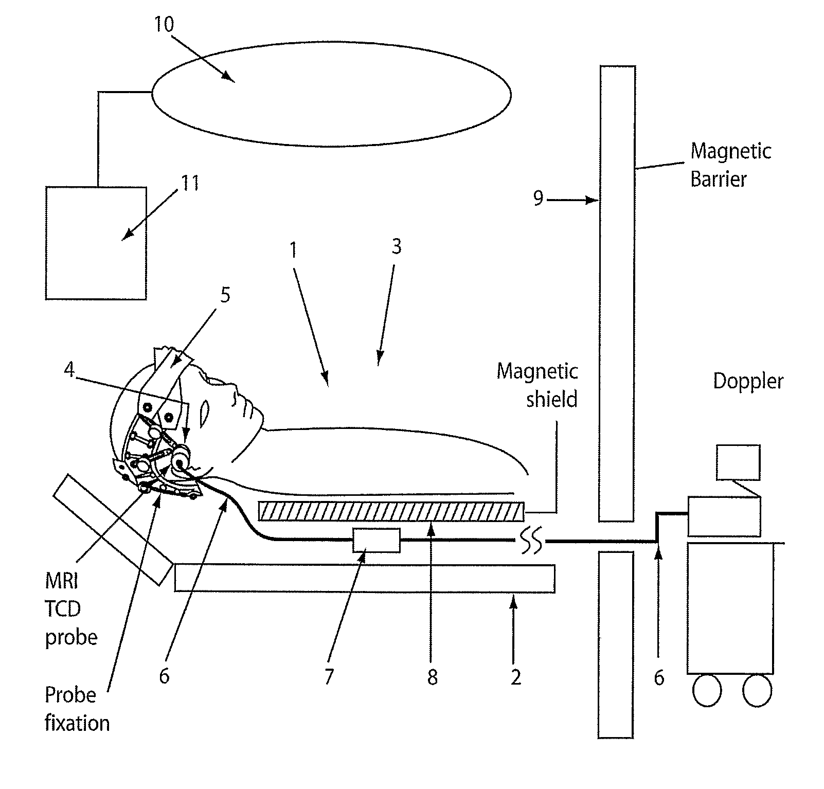
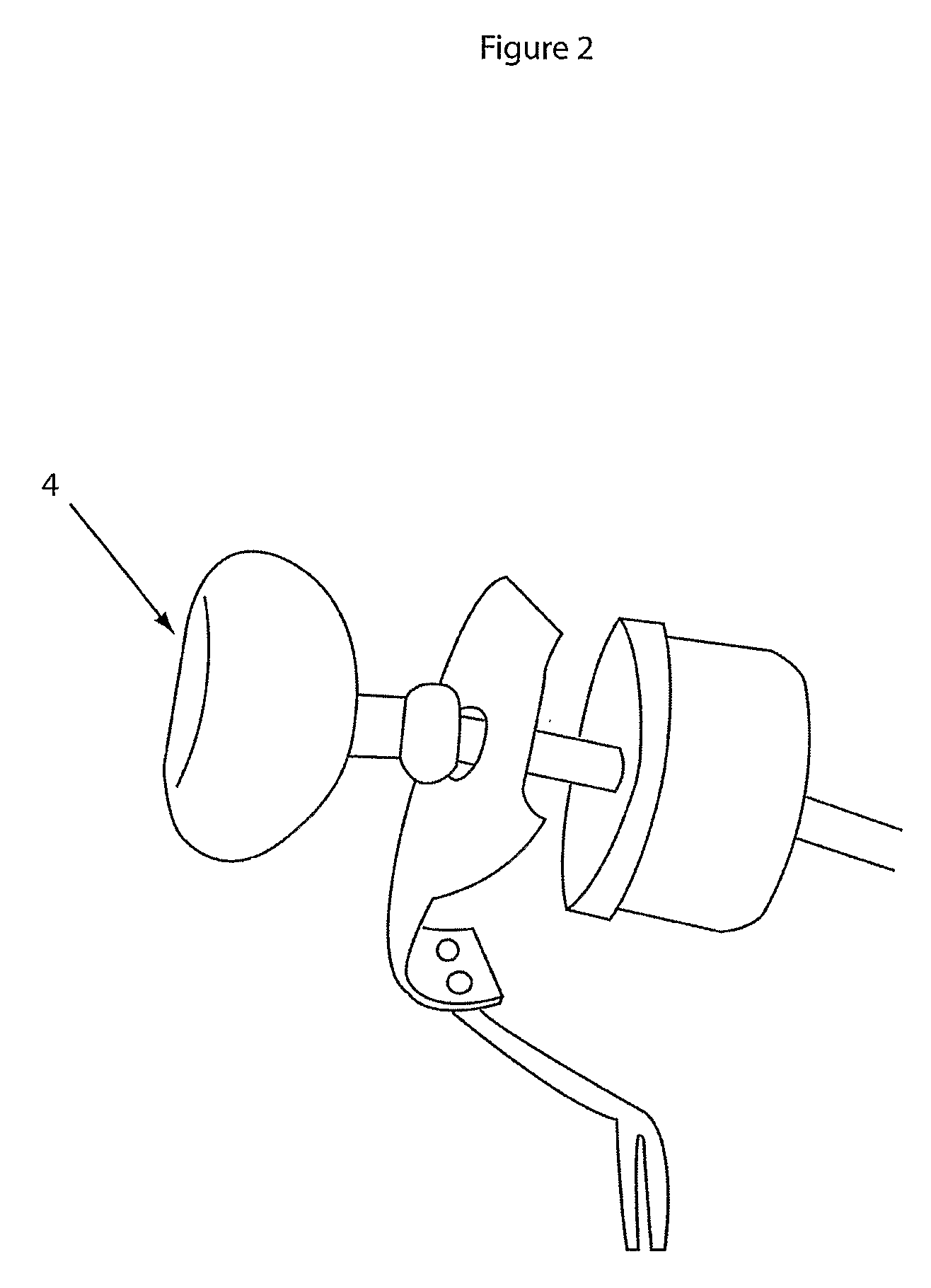
[0021]The invention is most easily understood with reference to the accompanying figures. FIG. 1 shows an embodiment of the invention, including the elements necessary to acquire ultrasound signals and spatial imaging information using strong magnetic fields. It will be understood that other embodiments of the invention are possible and that the scope of the invention is not limited to the embodiments described herein. In FIG. 1 is shown a subject 1 in a prone position on a table 2 within the magnetic field 3 of a spatial imaging device 10. The spatial imaging device may be any suitable spatial imaging device having a magnetic field. Preferably, the spatial imaging device is an MRI device. Other spatial imaging devices such as PET or CT devices are suitable for practising the invention. An ultrasound member 4 is engaged with a band 5 which, in turn, is engaged with the head 5 of the subject 1. Only one ultrasound member is shown in FIG. 1 but it is possible that more than one member...


PUM


Abstract

The present invention provides apparatus and methods for acquiring ultrasound measurements representing biosignals from a subject located in the strong magnetic field of a spatial imaging device. The invention includes ultrasound probes having minimal magnetic parts and an ultrasound signal pre-amplifier being shielded by a barrier or barriers from the strong magnetic field of a spatial imaging device such as an MRI, PET, or CT scanner. Most preferably, the ultrasound probe produces ultrasound waves in the frequency range of approximately 2.0 MHz to 2.5 MHz. The ultrasound probe may include no electronic components and contains minimal magnetic or ferromagnetic parts. The elements for acquiring and analysing ultrasound waves may be located in separate rooms from the imaging device. The invention provides a method of taking ultrasound measurements in quick succession with measurements of a spatial imaging device to provide a more informative understanding of the physiology of the subject.

Description

FIELD OF THE INVENTION[0001]This invention relates to methods and apparatus for measuring physiological parameters, in particular blood flow rates and electrophysiological activity, using ultrasound and spatial imaging techniques.BACKGROUND[0002]Developments in imaging techniques have made it possible to measure physiological parameters over differing timeframes by exploiting the physics of waveforms as they pass through heterogeneous tissues. For example, it is known in the art that the properties of the diffraction of sound waves of very high frequency (known as the Doppler effect) directed toward moving fluids, including blood moving through vessels, can be analysed to measure blood flow-rate. Such techniques using sound waves (known as ultrasound technologies) have been conveniently used in applications of cardiology and, more recently, brain blood-flow. Further developments in the use of ultrasound in brain blood-flow measurements are known in the art where measurements are mad...

Claims


Application Information

Patent Timeline
Patent Type & Authority Applications(United States)
IPC IPC(8): A61B5/055A61B8/00
CPCA61B5/6814A61B6/03A61B8/06A61B8/4227A61B8/0808A61B2019/5236G01R33/4814A61B6/4417A61B8/4416A61B5/0035A61B2090/374
Inventor WIDENHORN, GEROLD
Owner COMPUMEDICS
Why Eureka
Try Eureka