

Biomarkers and detection methods for gastric diseases
a gastric disease and biomarker technology, applied in the field of helicobacter pylori-related gastric diseases, can solve the problem of increasing the risk of non-cardiac g
- Summary
- Abstract
- Description
- Claims
- Application Information
AI Technical Summary
Benefits of technology
Problems solved by technology
Method used
Image

Examples
example 1
Identification of Gastric Cancer-Related Antigens of H. pylori
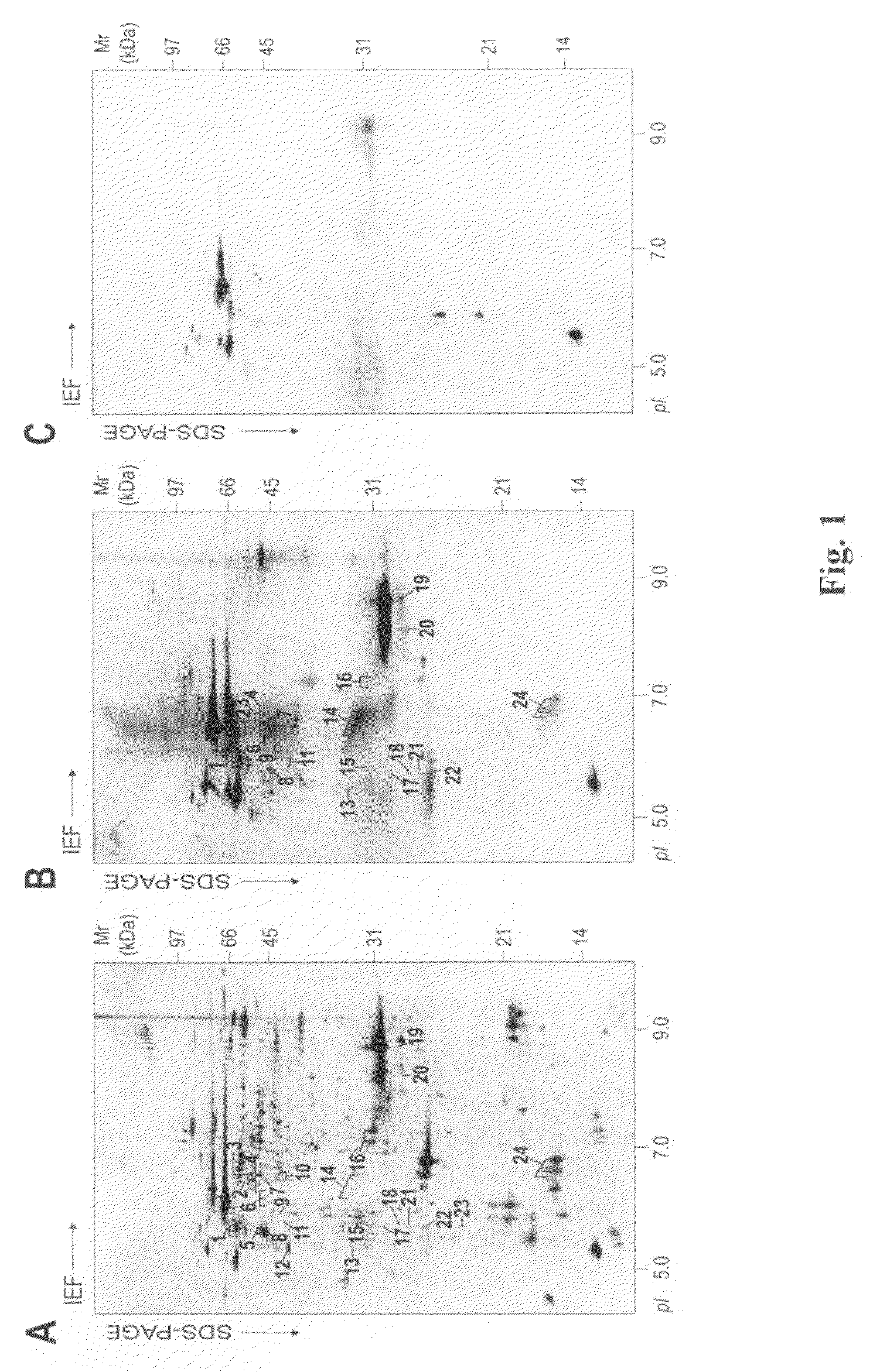
[0065]To identify candidate H. pylori antigens associated with GC, we performed 2D-SDS / PAGE on the bacterial proteins extracted with acidic glycine and compared the patterns of 2D-immunoblots probed with sera from H. pylori-infected patients with either GC or DU. Silver staining revealed a complex protein profile of the acid-glycine extract (FIG. 1A). Probing with 15 GC sera and 15 DU sera gave unique and different patterns of reactivity. Two representative immunoblots are shown in FIG. 1B (GC) and FIG. 1C (DU).
[0066]In general, the frequency of spot recognition was greater with GC sera than with DU sera. On the GC immunoblots, about 60 different reactive protein spots were detected, with molecular weights ranging from 14 to 85 kDa and pIs ranging from 4.5 to 9.5. Some of these antigenic spots were recognized by individual serum sample, but 49 spots were recognized by more than one. Comparing the antigenic protein profil...
example 2
GroES Seropositivity is Related to Gastric Cancer
[0069]To examine the clinicopathological significance of GroES seropositivity in H. pylori-infected patients, a GroES immunoblot assay was performed on a series of clinical samples. A serum was defined as GroES seropositive if rGroES was recognized by serum IgG. No seropositivity was seen with any serum sample from 32 healthy persons without H. pylori infection (controls). We then examined the serum IgG response to GroES in 313H. pylori-infected patients with GC (95 patients), gastritis (94 patients), or DU (124 patients). Overall, 42.8% of the H. pylori-infected patients gave a positive response. GroES seropositivity was related to patient age, increasing from 18.8% in patients aged less than 30 years to 40.2% inpatients aged 30-49 years (odds ratio (OR): 2.9, 95% confidence interval (CI): 0.8-10.9, P=0.1) and to 46.2% in patients aged more than 50 years (OR: 3.7, 956 CI: 1.0-13.4, P=0.04) (Supplemental Table III). Furthermore, the p...
example 3
Induction of Pro-Inflammatory Cytokine Production and COX-2 Expression in PBMC Stimulated with GroES
[0070]Example 2 demonstrated close association of GroES with GC, a cancer known to result from chronic inflammation caused by H. pylori infection. Moreover, GroES is a secreted protein and in direct contact with host, may mediate important interaction between H. pylori and host. We therefore investigated the effect of GroES on the inflammatory responses of mononuclear cells. PBMC were incubated with rGroES, then mRNA levels for 7 cytokines were determined by RT-PCR. As shown in FIG. 4A, rGroES stimulation caused a marked increase in IL-8, IL-6, IL-1β, and TNF-α, cytokines commonly found in H. pylori-infected patients. In addition, GM-CSF was slightly increased by rGroES (FIG. 4A), while IFN-γ and IL-12 were not changed (data not shown). Furthermore, mRNA levels of COX-2, an enzyme crucial for inflammatory responses, were also greatly enhanced after rGroES stimulation (FIG. 4A). These ...
PUM
| Property | Measurement | Unit |
|---|---|---|
| molecular weight | aaaaa | aaaaa |
| pH | aaaaa | aaaaa |
| pH | aaaaa | aaaaa |
Abstract
Description
Claims
Application Information