Histotripsy for thrombolysis

a thrombolysis and histopathology technology, applied in the field of ultrasonography, can solve the problems of increasing bleeding complications, affecting the normal formation of clots, and affecting the normal clotting of hemostasis, and achieves the effects of facilitating clotting, facilitating clotting, and reducing bleeding complications

Active Publication Date: 2009-07-09
RGT UNIV OF MICHIGAN
View PDF70 Cites 58 Cited by
  • Summary
  • Abstract
  • Description
  • Claims
  • Application Information

AI Technical Summary

Problems solved by technology

These thrombi can break off from site of formation and travel to distant sites (embolisation) and cause symptoms at sites distinct from the site of formation.
Each of these conditions poses a significant clinical problem.
Inhibition of normal hemostatic clot formation is associated with an increase in bleeding complications, which may be fatal in a small number of cases.
However, catheter-based devices are invasive and carry an increased risk of bleeding, damage to the vessel wall, and infection.
In rare cases, catheter-based thrombolysis methods may also result in death.
Direct surgical methods are even more invasive than catheter-based methods.
Even though this method increases thrombolysis efficiency, it still carries the undesired side effects of thrombolytic drugs.
This hybrid technique is still being studied and not currently in clinical use.
However, the mechanism is not well understood, and therefore, these techniques remain far from clinical application.
Specifically, when an acoustic field is propagated into a fluid, the stress induced by the negative pressure produced can cause the liquid to rupture, forming a void in the fluid which may contain vapor and / or gas.
However, no one has previously succeeded in achieving controlled and predictable cavitation for thrombolysis with real-time ultrasound feedback.

Method used


Image

  • Histotripsy for thrombolysis
  • Histotripsy for thrombolysis
  • Histotripsy for thrombolysis

Examples


Embodiment Construction

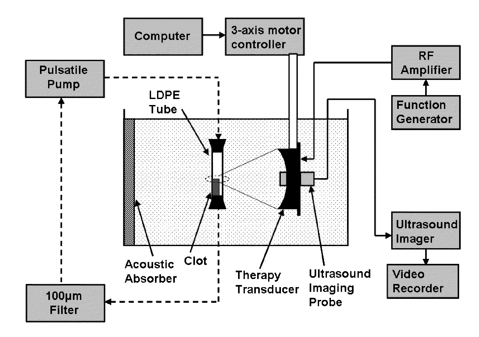
[0028]Example embodiments will now be described more fully with reference to the accompanying drawings.

[0029]Thrombosis is the formation of a blood clot in vasculature and is the primary cause of many vascular diseases, including heart attack, stroke, pulmonary embolism (PE) and deep vein thrombosis (DVT). Current clinical methods to treat thrombosis include anticoagulant and thrombolytic drugs, catheter-based surgical techniques, or a combination of the two where a catheter is used to locally deliver the thrombolytic agent to the site of occlusion. Thrombolytic drugs (e.g. Streptokinase, urokinase, rt-PA etc) administered without a catheter require long treatment times (several hours) and are non-specific, with a substantial risk of major bleeding that can be fatal in a small number of cases. The current catheter-based thrombolysis procedures include local delivery of thrombolytic agents by catheter, vein segment isolation and thrombolysis, and mechanical disruption and aspiration ...


PUM


Abstract

Methods for performing non-invasive thrombolysis with ultrasound using, in some embodiments, one or more ultrasound transducers to focus or place a high intensity ultrasound beam onto a blood clot (thrombus) or other vascular inclusion or occlusion (e.g., clot in the dialysis graft, deep vein thrombosis, superficial vein thrombosis, arterial embolus, bypass graft thrombosis or embolization, pulmonary embolus) which would be ablated (eroded, mechanically fractionated, liquefied, or dissolved) by ultrasound energy. The process can employ one or more mechanisms, such as of cavitational, sonochemical, mechanical fractionation, or thermal processes depending on the acoustic parameters selected. This general process, including the examples of application set forth herein, is henceforth referred to as “Thrombolysis.”

Description

CROSS-REFERENCE TO RELATED APPLICATIONS[0001]This application is a continuation-in-part of U.S. patent application Ser. No. 12 / 121,001 filed on May 15, 2008, which is a continuation-in-part of U.S. patent application Ser. No. 11 / 523,201 filed on Sep. 19, 2006, which claims the benefit of U.S. Provisional Patent Application No. 60 / 786,322, filed Mar. 27, 2006, U.S. Provisional Patent Application No. 60 / 719,703, filed Sep. 22, 2005, and U.S. Provisional Patent Application No. 60 / 753,376, filed Dec. 22, 2005. The U.S. patent application Ser. No. 12 / 121,001 filed on May 15, 2008 further claims the benefit of U.S. Provisional Patent Application No. 60 / 938,806, filed May 18, 2007. The present application further claims the benefit of U.S. Provisional Application No. 61 / 023,554 filed on Jan. 25, 2008. The entire disclosure of each of the above applications is incorporated herein by reference.FIELD[0002]The present teachings relate to ultrasound therapy and, more particularly, relate to met...

Claims


Application Information

Patent Timeline
Patent Type & Authority Applications(United States)
IPC IPC(8): A61B8/12A61B17/22
CPCA61B5/0536A61M37/0092A61B8/00A61B8/08A61B8/485A61B17/22004A61B17/2258A61B2017/00154A61B2017/00172A61B2017/00778A61B2017/22001A61B2017/22008A61B2017/22088A61B2017/22089A61B2019/5276A61B5/055A61B2090/378
Inventor MAXWELL, ADAMXU, ZHENGURM, HITINDER S.CAIN, CHARLES A.
Owner RGT UNIV OF MICHIGAN
Why Eureka
Try Eureka