Apparatus, a method and software for analyzing a cell image

Active Publication Date: 2009-08-06
EVIDENT CORP
View PDF2 Cites 19 Cited by
  • Summary
  • Abstract
  • Description
  • Claims
  • Application Information

AI Technical Summary

Benefits of technology

[0014]According to this structure, the judgment of a membrane translocation reaction is carried out without being influenced by a subjective judgment of an observer or an experimenter, and thereby, the reliability of the judgment result will increase. Moreover, since the operation of the above-mentioned apparatus can be automated through the image processing technique, it becomes unnecessary for an observer or an experimenter to make a judgment of a membrane translocation reaction in each measurement, which enables the reduction of the time and burden required for a judgment of a membrane translocation reaction.
[0015]In the above-mentioned structure, the fluorescence microscopic image may include fluorescence microscopic images of a plurality of cells (namely, a plurality of cells may be included in one image data), and in that case, the means that determines a region occupied by a cell may determine a region occupied by each of the plurality of cells; the means that determines a contour line of a cell may determine a contour line of each of the plurality of cells; and the means that judges a presence or an absence of

Problems solved by technology

Further, although no enormous labor would be required in judging the presence or absence of a membrane translocation reaction for one cell by means of the graph of fluorescence intensity distribution, the judging one by one for the increased number of cells requires much time and much burden.
Also, the variation in t

Method used


Image

  • Apparatus, a method and software for analyzing a cell image
  • Apparatus, a method and software for analyzing a cell image
  • Apparatus, a method and software for analyzing a cell image

Examples


Embodiment Construction

[0032]In the followings, the present invention is explained in detail about some preferable embodiments, referring to the attached drawings.

Structure of a Fluorescence Microscope Observation System

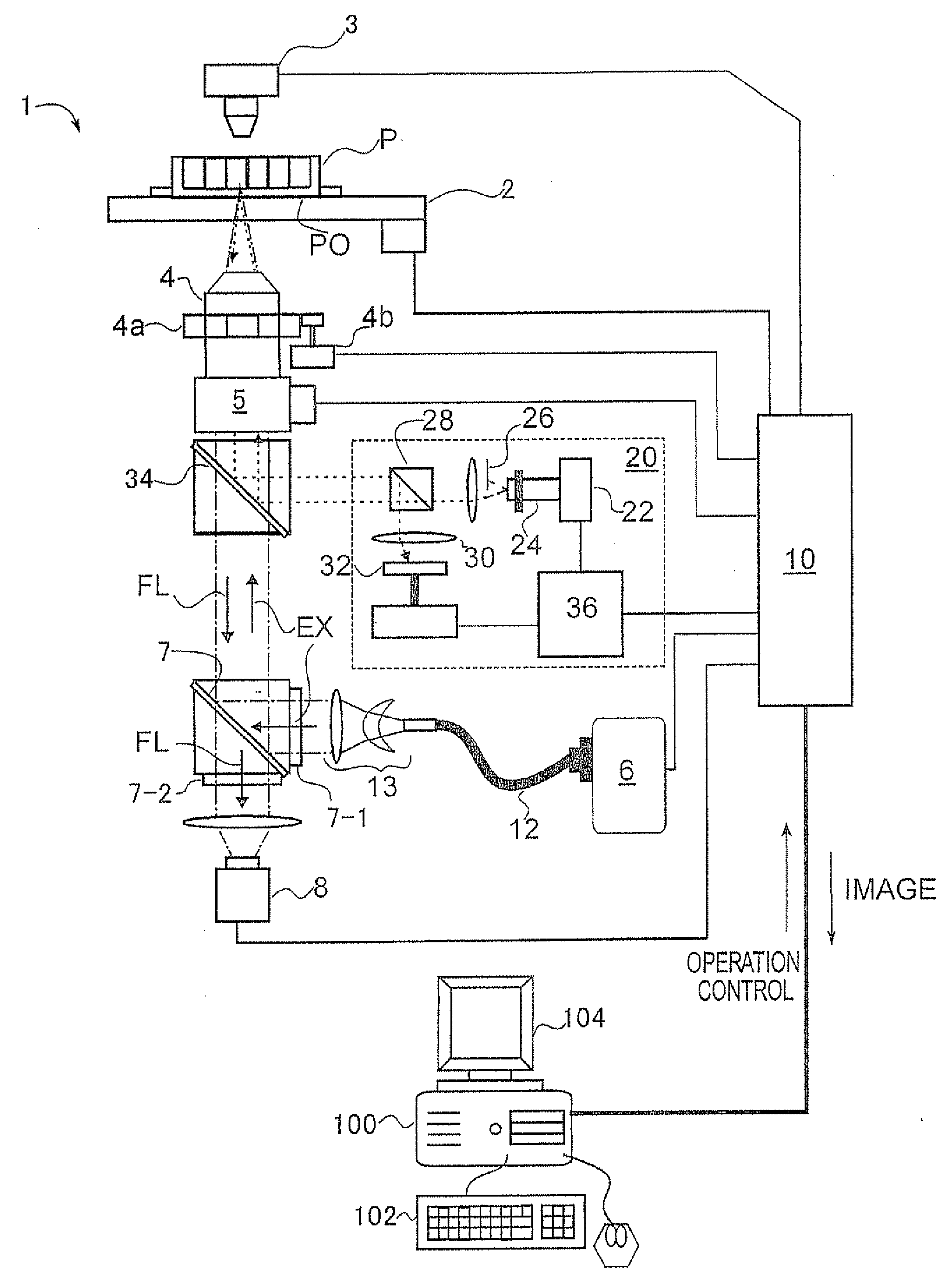
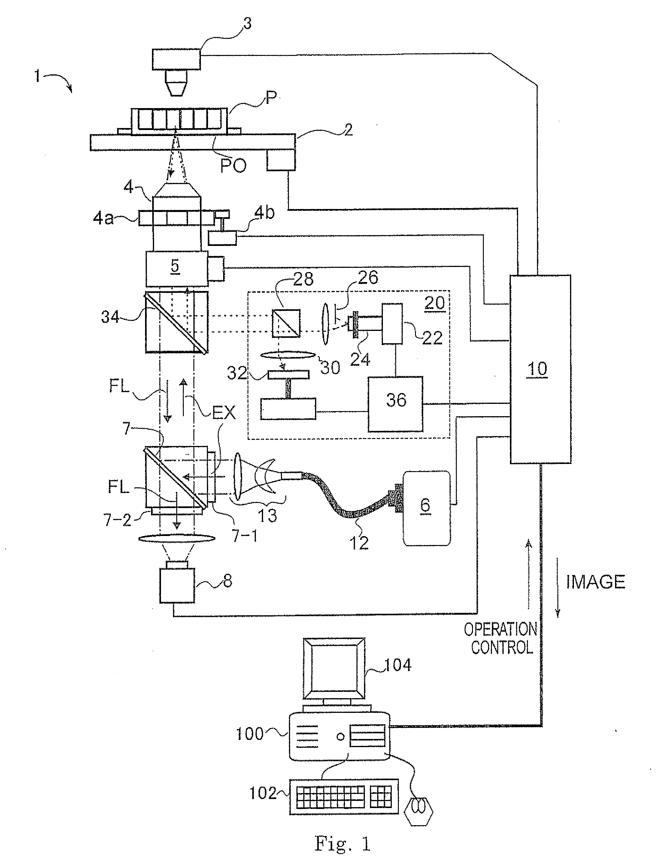
[0033]FIG. 1 schematically represents a fluorescence microscope observation system incorporating a preferable embodiment of a cell image analysis apparatus for the judging of a membrane translocation reaction of a cell in accordance with the present invention. In the drawing, portions, not relevant to the structures of the present invention, are omitted for the simplification of the explanation.

[0034]Referring to the drawing, the fluorescence microscope observation system includes an inverted type fluorescence microscope 1 and an image analysis apparatus 100. Similarly to normal fluorescence microscopes, the fluorescence microscope 1 comprises a stage 2 electrically movable in the XY directions (horizontal) on which stage a cell sample P (for example, plastic micro titer plate having 24 we...


PUM


Abstract

There are provided a cell image analysis apparatus or method for judging the presence or absence of a membrane translocation reaction quickly and with good reproducibility using a fluorescence microscope. The inventive apparatus is a cell image analysis apparatus for judging the presence or absence of a membrane translocation reaction occurring in a cell owing to application of an arbitrary stimulus to the cell, characterized by comprising: a means that acquires as an image data a fluorescence microscopic image of a cell after the application of the stimulus; a means that determines a region occupied by the cell in the fluorescence image of the cell; a means that determines a contour line of the cell which has a width of a predetermined number of pixels from a periphery of the region occupied by the cell; and a means that judges the presence or absence of the membrane translocation reaction based on a brightness value on the contour line of the cell and a brightness value in an inside of the contour line of the cell.

Description

TECHNICAL FIELD[0001]This invention relates to an apparatus and a method of an image analysis of a microscopic image of a cell obtained with an optical microscope, and software for them, and more particularly, to an apparatus, a method and software for analyzing a fluorescence microscopic image of a cell to judge the presence or absence of occurrence of a membrane translocation reaction observed in a cell.BACKGROUND ART[0002]It is known that, in various cells, chemical stimulation with a bioactive substance or a drug, or physical stimulation with light, electricity, mechanical force, etc, induces a phenomenon, called as “membrane translocation reaction”, of accumulation of certain kinds of intracellular proteins onto a cell membrane. As well known in ones skilled in the art, when a membrane receptor, such as a G protein binding protein on a cell membrane (GPCR: G Protein Coupled Receptor), receives a specific substance (ligand) or a photon, etc. from the exterior, this becomes a tri...

Claims


Application Information

Patent Timeline
IPC IPC(8): G06K9/00
CPCG01N21/6458G06K9/00127G06T2207/30024G06T2207/10056G06T2207/10064G06T7/0012G06V20/69
Inventor KOBAYASHI, TAMIYOTAKAGI, KOSUKEMATSUO, YUICHIRO
Owner EVIDENT CORP
Why Eureka
Try Eureka