Method and device for examining a biological tissue

a biological tissue and method technology, applied in the field of biological tissue methods and devices, can solve the problems of insufficient precision in detecting fluorescent light from deep tissue layers, tumors or lesions located deep in the tissue cannot be detected safely and reliably, and the local resolution of the method can be further improved, and the local resolution can be simplified.

Inactive Publication Date: 2009-08-27
SIEMENS AG
View PDF4 Cites 6 Cited by
  • Summary
  • Abstract
  • Description
  • Claims
  • Application Information

AI Technical Summary

Benefits of technology

[0021]According to one embodiment of the invention, a gradient field is used as a magnetic field. A modification of the permutation symmetry imbalance occurs when an alternating field that fulfils the known condition of resonance is irradiated. The condition of resonance is fulfilled, for example, if the frequency of the alternating field corresponds to the Larmor frequency of spins in the magnetic field. In a gradient field, the condition of resonance is a function of the location in the tissue. As a result of the size of the gradient field and the gradient intensity being known, an alternating field that fulfils the condition of resonance at a given location can be radiated into the tissue. The detection of the luminescent light or modification thereof can be simplified depending on the location.
[0022]To generate the magnetic field or the gradient field and the alternating field, it is possible to use magnetic coils and / or permanent magnets. The alternating field is preferably generated by means of magnetic coils. The alternating field can be radiated into the tissue in such a way that it acts upon the entire tissue. It is also possible, however, to radiate the alternating field only onto a given limited area located in the tissue. The position of the area can also be used as additional local information to improve the local resolution of the method.
[0023]A further embodiment of the invention makes provision for generation, modification, irradiation and / or detection means inserted into a cavity located in the tissue or leading to the tissue can be used to generate and / or modify the permutation symmetry imbalance and / or to irradiate the tissue and / or detect luminescence. This allows an examination of the tissue using a minimally invasive method, for example, via a vein, the trachea or the intestine. The generation, modification, irradiation and / or detection means can be inserted into the cavity by using a catheter, a probe or such like, for example. Using incoming lines, outgoing lines, control lines and suchlike connected to the generation, modification, irradiation and / or detection means, the function and / or movement thereof in the cavity can be controlled manually or even automatically. The generation, modification, irradiation and / or detection means can also be provided as units that are separate from one another or combined in any desired combinations. The generation, modification, irradiation and / or detection means can be combined in particular into a single unit configured along the lines of a probe that can be inserted into the tissue. Generation, modification, irradiation and / or detection means configured in such a way can be brought via the cavity essentially directly to the location of the tissue that is to be examined. The local resolution can be further improved. The tissue can be examined with even greater precision.
[0024]According to one embodiment of the invention, a light conductor is used to conduct the electromagnetic radiation and / or the luminescent light. A light conductor can be inserted into the cavity via a catheter or a probe. The electromagnetic radiation can thus be directed straight to the tissue. Likewise, the luminescent light generated in the tissue can be directed to the detection means using a light conductor. Absorption and scatter losses in or outside the tissue can be reduced. Even more precise results can be achieved in the examination.

Problems solved by technology

A disadvantage thereof is that tumors or lesions located deep in the tissue cannot be detected safely and reliably due to the blurring caused by the scattering of the light in the tissue.
As a result of the scattering of the fluorescent light in the tissue, it is not possible to detect fluorescent light from deep layers of tissue with sufficient precision.
Tumors or lesions located deep in the tissue cannot be detected safely and reliably.
A disadvantage of the method is the limited local resolution caused by the extensive light scattering in the tissue.
As a result thereof, the achievable local resolution of the tomographic images is limited.
The acoustic connection between a receiver and the tissue that is required to carry out the photoacoustic tomographic method is expensive.
It is possible, however, that where there is a poor contrast, small lesions, tumors or suchlike cannot be detected precisely and reliably.

Method used


Image

  • Method and device for examining a biological tissue
  • Method and device for examining a biological tissue
  • Method and device for examining a biological tissue

Examples


Embodiment Construction

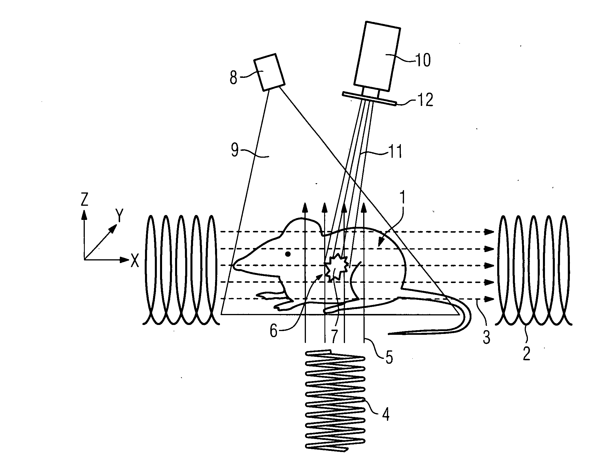
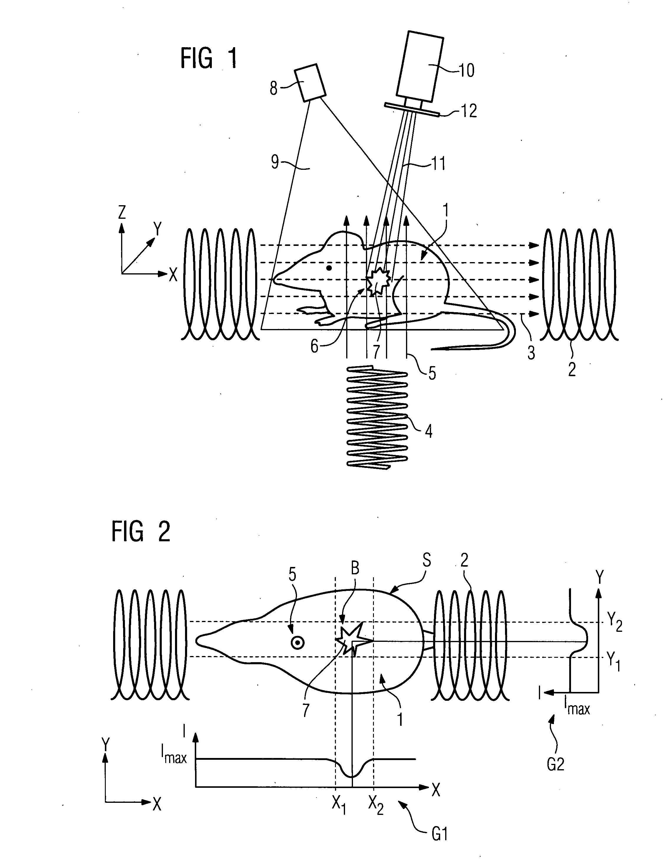
[0035]A magnetic field 3 is generated in a tissue 1 of a mouse, using two first magnetic coils 2, for example. A second magnetic coil 4 is used to generate a magnetic alternating field 5 essentially perpendicular to the magnetic field 3. A luminescent substance 7 is accumulated in a tumor 6 located in the tissue 1. An excitation light emanating from a light source 8 to excite the luminescent substance 7 is denoted by the reference sign 9. The reference sign 10 denotes a CCD camera for detecting a luminescent light 11 emanating from the luminescent substance 7. A filter 12 is connected upstream of the CCD camera. X, Y and Z are used to denote an X, Y and Z direction. X1 and X2 and Y1 and Y2 denote first and second X and Y co-ordinates respectively.

[0036]The method is implemented as follows:

[0037]In a first step, a permutation symmetry imbalance is generated in the tissue 1 of the mouse using the magnetic field 3 generated using the first magnetic coils 2. The permutation symmetry imb...


PUM


Abstract

The invention relates to a method and a device for analyzing a biological tissue, whereby a luminescence light of a luminescence substance is detected. The aim of the invention is to increase the precision and reliability of the analysis. To this end, a permutation symmetry imbalance is generated in the tissue by a magnetic field, the permutation symmetry imbalance is modified at a pre-determined location by a magnetic alternating field, and the luminescence light is detected according to the pre-determined location.

Description

CROSS REFERENCE TO RELATED APPLICATIONS[0001]This application is the US National Stage of International Application No. PCT / EP2006 / 061577 filed Apr. 13, 2006 and claims the benefits thereof. The International Application claims the benefits of German application No. 10 2005 017 817.0 filed Apr. 18, 2005, both of the applications are incorporated by reference herein in their entirety.FIELD OF THE INVENTION[0002]The invention relates to a method and a device for examining a biological tissue.BACKGROUND OF THE INVENTION[0003]Umar Mahmood et al., “Near Infrared Optical Imaging of Protease Activity for Tumor Detection”, Radiology 1999, 213: 866-870, discloses a method and a device for detecting tumors in mice. To detect the tumor, light is detected by a fluorogen that is activated by tumor proteins. A disadvantage thereof is that tumors or lesions located deep in the tissue cannot be detected safely and reliably due to the blurring caused by the scattering of the light in the tissue.[000...

Claims


Application Information

Patent Timeline
Patent Type & Authority Applications(United States)
IPC IPC(8): A61B5/055G01R33/32
CPCA61B5/055G01R33/4808G01N24/10
Inventor BOHME, CHRISTOPHHOHEISEL, MARTINLIPS, KLAUSPFISTER, MARCUS
Owner SIEMENS AG
Why Eureka
Try Eureka