
Method For Identification Of Novel Physical Linkage Of Genomic Sequences

a genomic sequence and physical linkage technology, applied in the field of genomic sequence physical linkage identification, can solve the problems of difficult or impossible sequence analysis of nonfixed or copy number variable elements in the genome, not applying to other strains of the same species, and affecting the identification speed and economic

Inactive Publication Date: 2009-10-22
RUTGERS THE STATE UNIV +2
View PDF18 Cites 8 Cited by
  • Summary
  • Abstract
  • Description
  • Claims
  • Application Information

AI Technical Summary

Benefits of technology

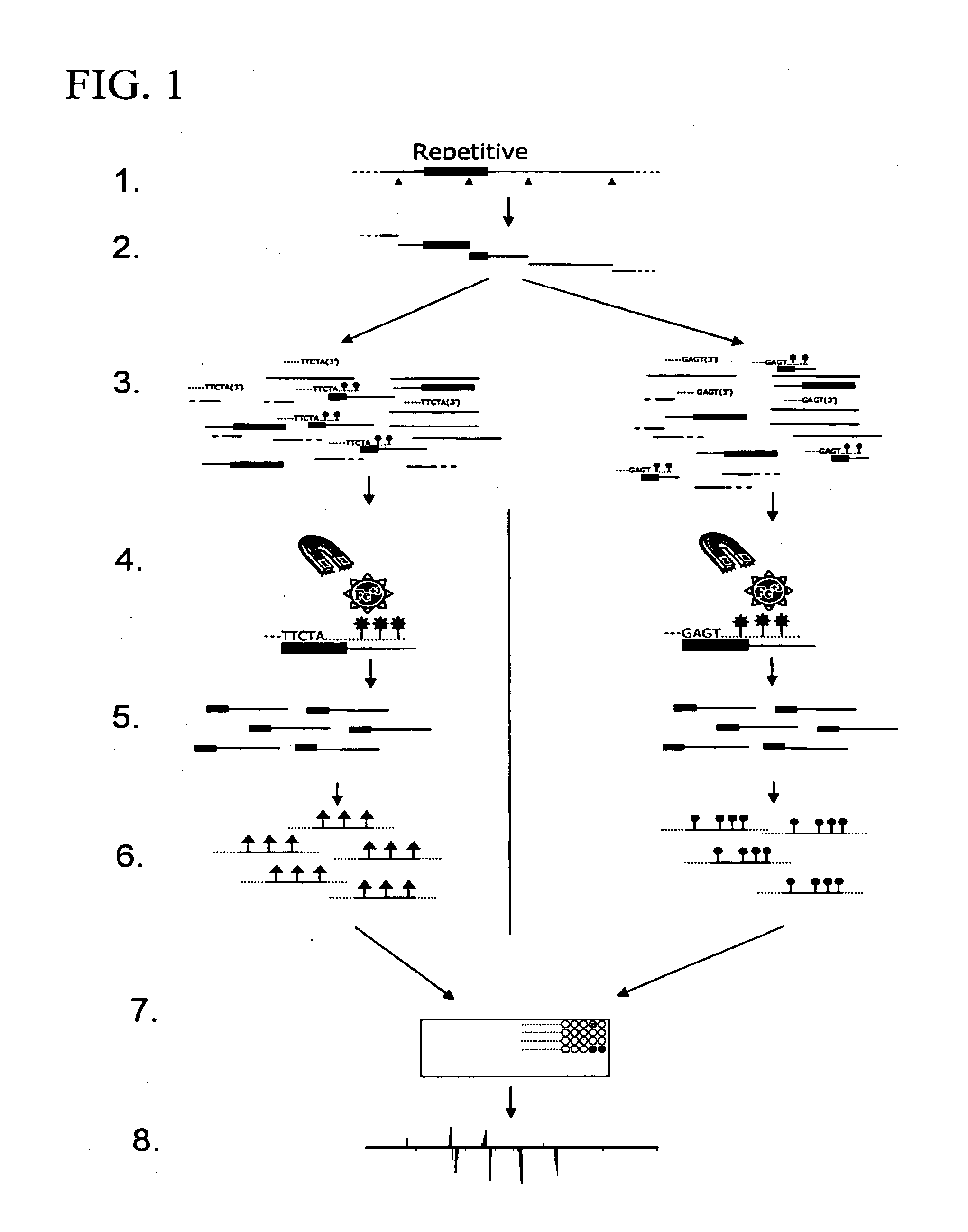
[0014]The invention is based on the discovery of a method for rapidly and economically identifying the location in a genome of a nonfixed or multicopy genomic element of interest. The method involves isolating a genomic nucleic acid fragment that contains the genomic element and a flanking sequence from the genome, labeling the isolated fragment to form a labeled probe, and applying the labeled probe to a sufficiently dense genomic microarray such that specific binding of the probe to one or more positions on the microarray can be determined and thus the location of the genomic element of interest can be determined. Alternatively, the labeling of the isolated fragments may occur after immobilization as part of a sequencing process, such as by successively attaching individual nucleotides to template fragments on a surface and thereby determining their sequence.

Problems solved by technology

However molecular analysis to determine the positions of nonfixed or copy number variable elements throughout the genome can be difficult or impossible to determine by sequence analysis due to the problem of properly assembling relatively short reads generated by random shotgun sequencing into their proper genomic context of potentially much larger repetitive elements, segmental duplications, translocations, inversions or other chromosomal rearrangements.
This has become an acute problem for so-called “next-generation sequencing (NGS)” approaches that rely on the genome wide assembly of very short read lengths (typically 10-30 base pairs, sometimes 30-100 base pairs), especially in combination with more complex genomes, such as the human genome.
While whole genome sequencing can identify all transposable or multicopy elements in the specific genome under examination, the results may not apply to other strains of the same species.
However, the identification of insertion sites remains a methodological challenge in insertional mutagenesis.
The challenge for identifying the location of nonfixed, multicopy or randomly inserted genomic elements is the identification of the sequences which flank these genomic elements.
Although laborious and expensive, sequencing of cloned or PCR-amplified flanking fragments unequivocally identifies insertion sites, and databases of insertion-site sequences have been established for some genomes.
However, all of these methods suffer from either a limitation that they permit screening for insertions in only one or a small number of genes at a time, or require use of semispecific PCR, which can be expensive, time-consuming, biased and incomplete.
Likewise, the proper assembly of genomic sequences that contain copy number variants or other rearrangements can be very difficult.
The detection of gross chromosomal rearrangements in the genome of patients with genetic diseases by oligonucleotide microarrays or fluorescence in situ hybridization (FISH) is cumbersome and typically limited to a region of about 10-20 kilobases near a breakpoint.
The routine assembly of larger blocks of contiguous, intergenic haplotype information from individual samples has been unattainable using current systems, and no solutions exist to deconvolute complex genomic regions related to copy number variations, repetitive elements and segmental duplications in a high-throughput mode.

Method used


Image

  • Method For Identification Of Novel Physical Linkage Of Genomic Sequences
  • Method For Identification Of Novel Physical Linkage Of Genomic Sequences
  • Method For Identification Of Novel Physical Linkage Of Genomic Sequences

Examples


example 1

Introduction

[0078]The model eukaryote S. cerevisiae has been at the forefront of studies of retrotransposons, i.e. transposons that use reverse transcriptase for their replication, and which copy and paste themselves to new genomic locations. Several distinct families of retrotransposons, or “Tys” have been identified in this organism, both anecdotally, and systematically through the genome sequencing effort. In the only fully sequenced S. cerevisiae strain, S288c, the most abundant transposons are Ty1 (31 copies) and Ty2 (11 copies). These closely related 5.9 kb full-length mobile elements consist of two overlapping open reading frames, each of which encodes several proteins. The coding regions are flanked by ˜300 bp nearly identical long terminal repeats (LTRs). Ty4 (3 copies) is a distinct and less abundant element with a similar structure. Ty3 (2 copies) is another distinct element, with a different arrangement of protein coding segments, but still with flanking LTRs. Ty5 is onl...

example 2

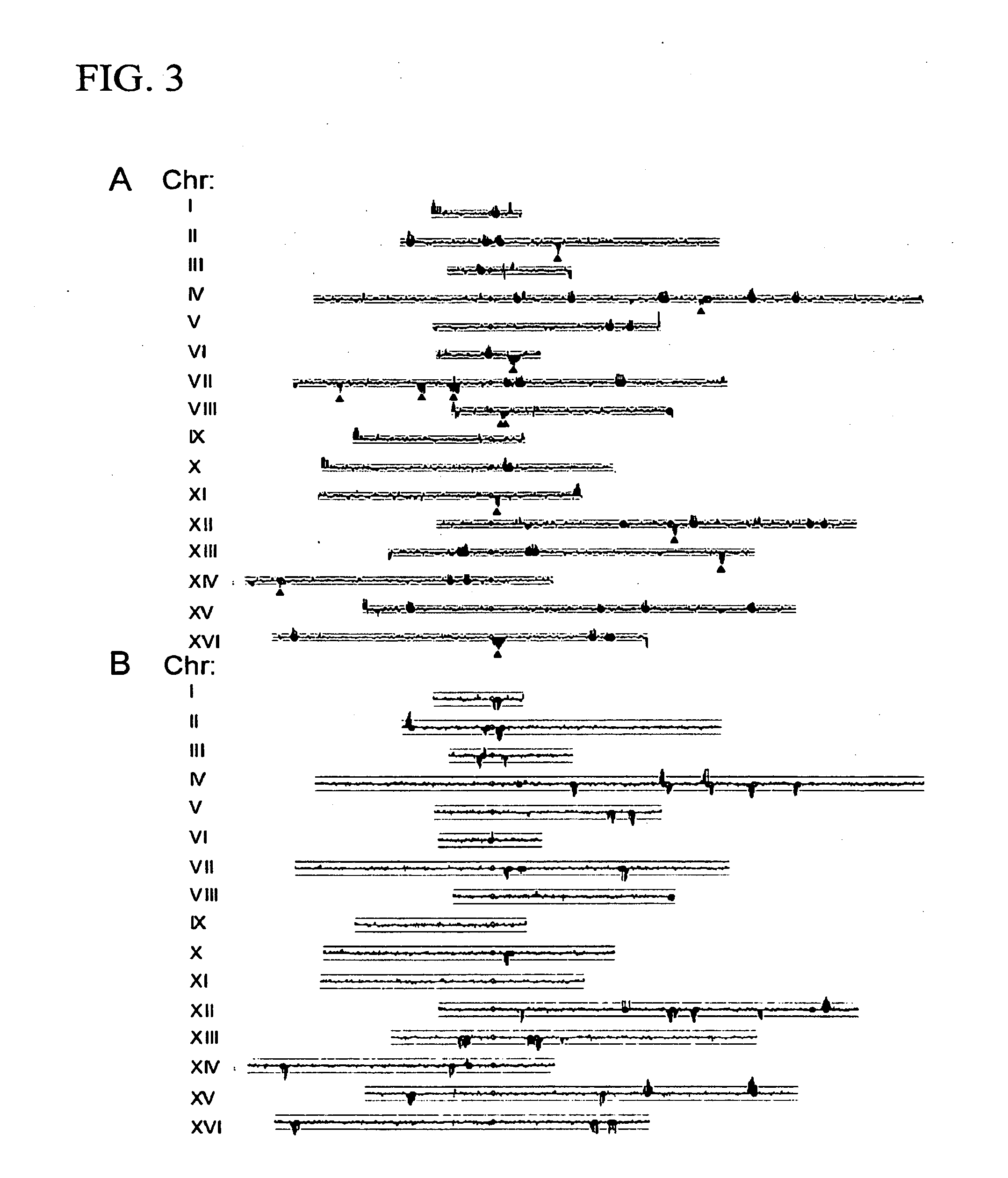
[0096]The following example shows how the method of the present invention can be used to extract and identify DNA associated with any specific sequence. In particular, probes were designed that would anneal to internal regions of Ty1 or Ty2, exploiting the regions of maximum differences between these two families of closely related elements. As shown in FIG. 4, when Ty1-associated fragments were labeled with Cy3 and Ty2-associated fragments with Cy5, each initial Ty1 / 2 peak could be correlated with the respective element associated with it. We extended this analysis to identify the three Ty3 full-length elements and Ty3 and Ty4 solo LTR elements in the S288c genome.

example 3

[0097]The following example shows how the method of the present invention can be extended to partially unmapped strains.

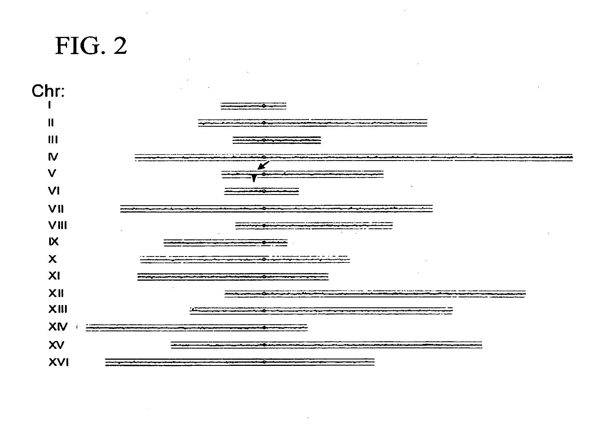
[0098]A comparison was made in the pattern of transposons in S288c with those in two common lab strains, CenPK and W303. In each of these cases, the strain was originally derived from a cross between S288c and an unrelated strain, although the detailed histories and origins are not completely documented. Previous work has shown that these strains are patchworks, with blocks of S288c sequence interspersed with blocks from the other parent (Daran-Lapujade et al., 2003; Winzeler et al., 2003). Using Affymetrix yeast tiling arrays, which are based on the S288c sequence, the patchwork nature of these strains is easily observable (FIG. 4), since SNPs are much more likely to be present and detected for segments derived from the non-S288c parent. We took advantage of this analysis to align each S288c, W303, and CenPK chromosome with the respective chromosome tracing derive...


PUM

PropertyMeasurementUnit
Fluorescenceaaaaaaaaaa

Abstract

The invention is directed to methods to identify the location in a genome of a nonfixed or multicopy genomic element using microarrays or sequencing.

Description

CROSS-REFERENCE TO RELATED APPLICATIONS[0001]This application claims the benefit of U.S. Provisional Application No. 60 / 800,426, filed May 15, 2006, and U.S. Provisional Application No. 60 / 833,042 filed Jul. 25, 2006, both of which are herein incorporated by reference in their entireties.STATEMENT REGARDING FEDERALLY SPONSORED RESEARCH[0002]The U.S. government may have certain rights in this invention as provided for by the terms of grants R44 AI 51036-02 and P50 GM071508, both awarded by the National Institutes of Health.FIELD OF THE INVENTION[0003]The present invention relates to methods for identifying the presence and location of nucleic acid segments within a genome.BACKGROUND OF THE INVENTION[0004]Whereas the location of most genomic sequences is fixed along a chromosome, some genomic elements are nonfixed or may occur in multiple copies. Nonfixed genomic elements, such as transposable elements, chromosomal rearrangement breakpoints, natural viral insertions, artificial insert...

Claims


Application Information

Patent Timeline
IPC IPC(8): C12Q1/68
CPCC12Q1/6874C12Q1/6813
Inventor DAPPRICH, JOHANNESGABRIEL, ABRAMDUNHAM, MAITREYA
Owner RUTGERS THE STATE UNIV
Learn More