
Magnetic immunohistochemical staining device and methods of use

a magnetic immunohistochemical and staining technology, applied in the field of immunology-based techniques, can solve the problems of microscope slides not being able to have a barrier prepared in advance, freezing sectioning is not always an optimal choice for researchers, etc., and achieve the effect of decreasing the amount of reagents needed

Inactive Publication Date: 2009-12-31
UNIV OF SOUTH FLORIDA
View PDF2 Cites 2 Cited by
  • Summary
  • Abstract
  • Description
  • Claims
  • Application Information

AI Technical Summary

Benefits of technology

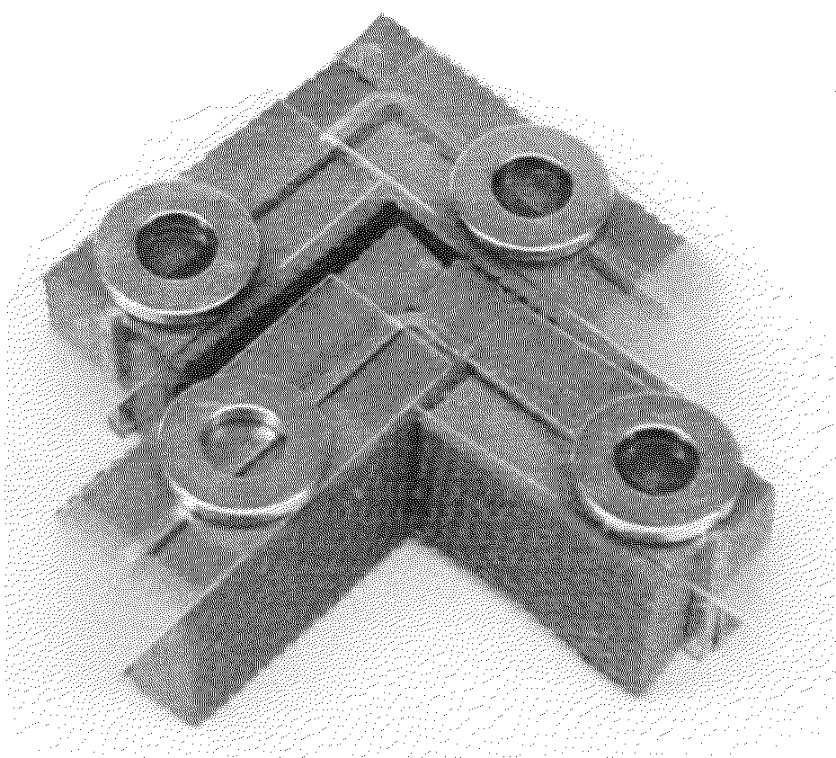
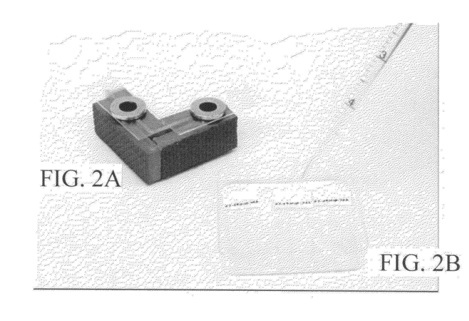
[0004]The subject invention concerns devices for staining a slide-mounted specimen of interest, such as tissue sections, cells, and the like. A device of the invention comprises a free-standing metal bracket. In one embodiment, the invention described herein is comprised of an L-shaped or V-shaped metal bracket with a plastic block or foam mat attached on one or both sides of the bracket, wherein a slide may be mounted with a tissue section of interest. The device further comprises a magnet placed on the device wherein the magnet has an opening to allow solutions to be added onto the specimen of interest, thereby decreasing the amount of reagents necessary to stain the specimen. Multiple devices can be placed together for multiple staining applications. An advantage of the subject device over staining platforms available in the art is that less reagent or staining solution is needed to achieve the same result as a standard staining application. For example, in one embodiment, only about 300 microliters of antibody solution is necessary to stain a specimen of interest with this device, compared to at least 4 milliliters of solution in other devices in the art (FIGS. 3-5). Moreover, it can be envisioned that other types of reagents of interest can be useful with this device to stain materials on a slide.

Problems solved by technology

Frozen sectioning is not always an optimal choice for researchers, since the tissue can distort while it is being cut, and since the sections are difficult to mount on slides when they are less than 15 microns thick.
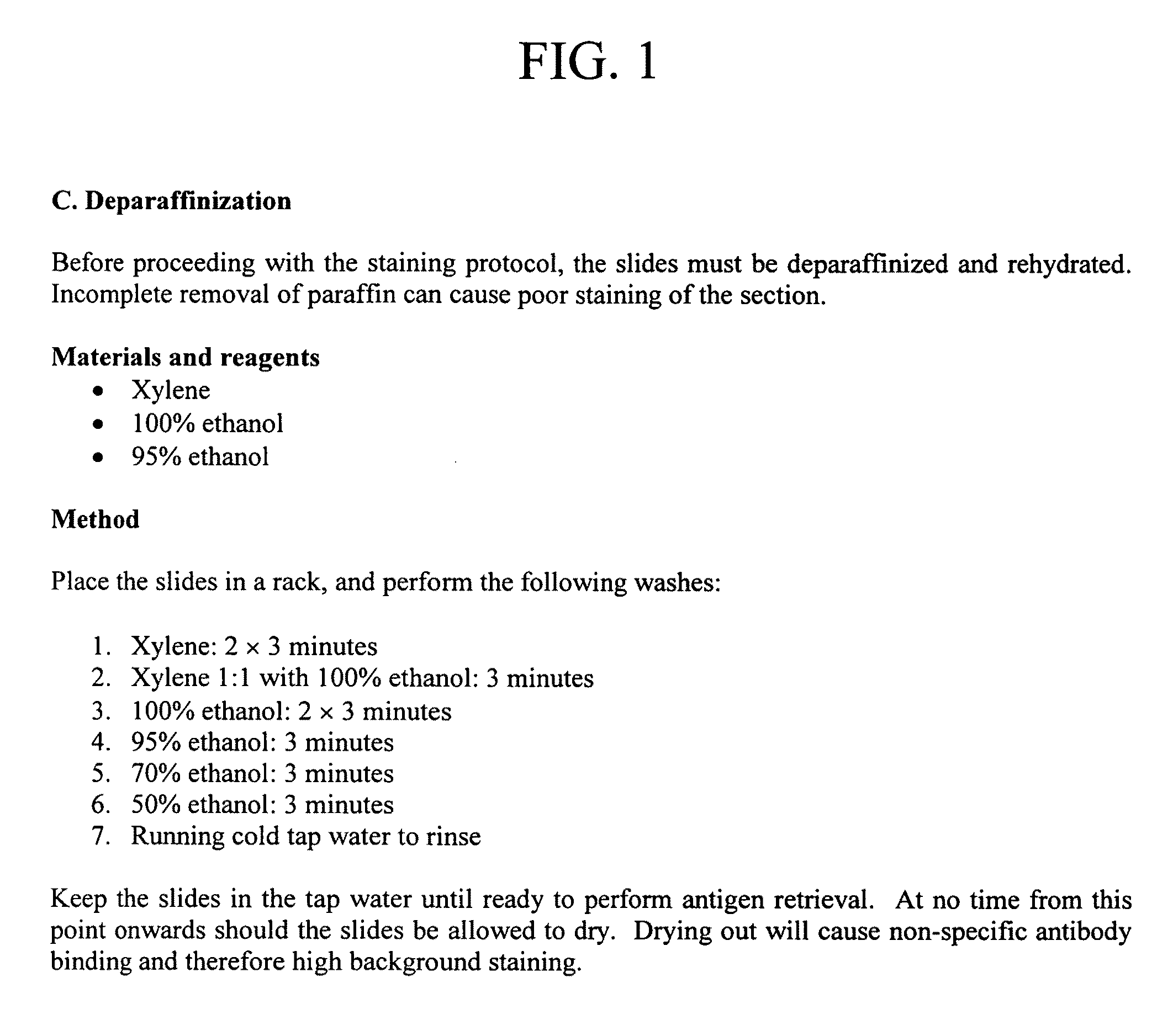
However, the microscope slides cannot have a barrier prepared in advance, due to the steps involved in deparaffinization, optional antigen retrieval, and followed immediately by IHC. FIG. 1 shows a standard deparaffinization protocol, written by ABCAM (a common antibody supplier), which also notes reasons why the tissue cannot dry out.

Method used


Image

  • Magnetic immunohistochemical staining device and methods of use
  • Magnetic immunohistochemical staining device and methods of use
  • Magnetic immunohistochemical staining device and methods of use

Examples


Embodiment Construction

[0020]The subject invention concerns a staining device for staining a slide-mounted specimen of interest, such as tissue sections, cells and the like. A device of the invention comprises a free-standing metal bracket. A metal bracket of the device, in one embodiment, can have a general L- or V-shape, although other similar shapes are contemplated within the scope of the invention (e.g., shapes where the angle formed by the bend of the bracket is greater than or less than 90°). In one embodiment (FIG. 10A), a staining device of the invention comprises a metal L-shaped bracket (10), a plastic block or foam mat (12) (e.g., “Foam Linking Mat” (Lowes Home Improvement, Item# 168520)), and a magnet (14) having an opening (15). The plastic block or foam mat (12) can be cut and glued to the metal bracket (10) with a silicon-based adhesive (e.g., DAP Auto / Marine Sealant), but alternative adhesives would also be appropriate. The adhered plastic block or foam mats (12) on both sides of the meta...


PUM

PropertyMeasurementUnit
distanceaaaaaaaaaa
thickaaaaaaaaaa
thicknessesaaaaaaaaaa

Abstract

The subject invention concerns a device for use in staining samples or tissues of interests on slides. The subject invention also concerns methods of using the device to stain a specimen on a slide or to detect antibody and proteins of interest of a tissue section.

Description

CROSS-REFERENCE TO RELATED APPLICATION[0001]The present application claims the benefit of U.S. Provisional Application Ser. No. 61 / 132,945, filed Jun. 24, 2008, which is hereby incorporated by reference herein in its entirety, including any figures, tables, nucleic acid sequences, amino acid sequences, and drawings.BACKGROUND OF THE INVENTION[0002]Immunohistochemistry (IHC) is an immunology-based technique that allows for the localization and presence of proteins within tissue sections. IHC protocols involve rehydrating slide-mounted tissue sections, washing and blocking unintended binding sites, and administering an antibody solution, under gentle agitation and time and temperature constraints, which allow the antibody to bind to specific target proteins within the tissue. Microscopy is later used to analyze the antibody-stained tissue sections.[0003]There are several methods for preparing and sectioning tissues so that they can be mounted upon slides. One method consists of sectio...

Claims


Application Information

Patent Timeline
Patent Type & Authority Applications(United States)
IPC IPC(8): G01N1/30G01N1/31
CPCG01N1/312
Inventor BOYD, TIMOTHY
Owner UNIV OF SOUTH FLORIDA
Learn More