Apparatus and method for indicating likely computer-detected false positives in medical imaging data

Active Publication Date: 2010-01-07
KONINKLJIJKE PHILIPS NV
View PDF11 Cites 9 Cited by
  • Summary
  • Abstract
  • Description
  • Claims
  • Application Information

AI Technical Summary

Benefits of technology

[0019]This provides the advantage of providing medical image data including computer-detected features of interest that are indicated by different graphical markers depending on whether the feature o

Problems solved by technology

However, false positive markings often occur close to the beating heart due to image artefacts caused by cardiac motion.
The heart beat causes erroneous displacements of the image slices, which in turn leads to an apparent discontinuity in, for example, vessel structures, airways and other anatomical features, which can be misinterpreted by the CAD software and thus

Method used


Image

  • Apparatus and method for indicating likely computer-detected false positives in medical imaging data
  • Apparatus and method for indicating likely computer-detected false positives in medical imaging data
  • Apparatus and method for indicating likely computer-detected false positives in medical imaging data

Examples


Embodiment Construction

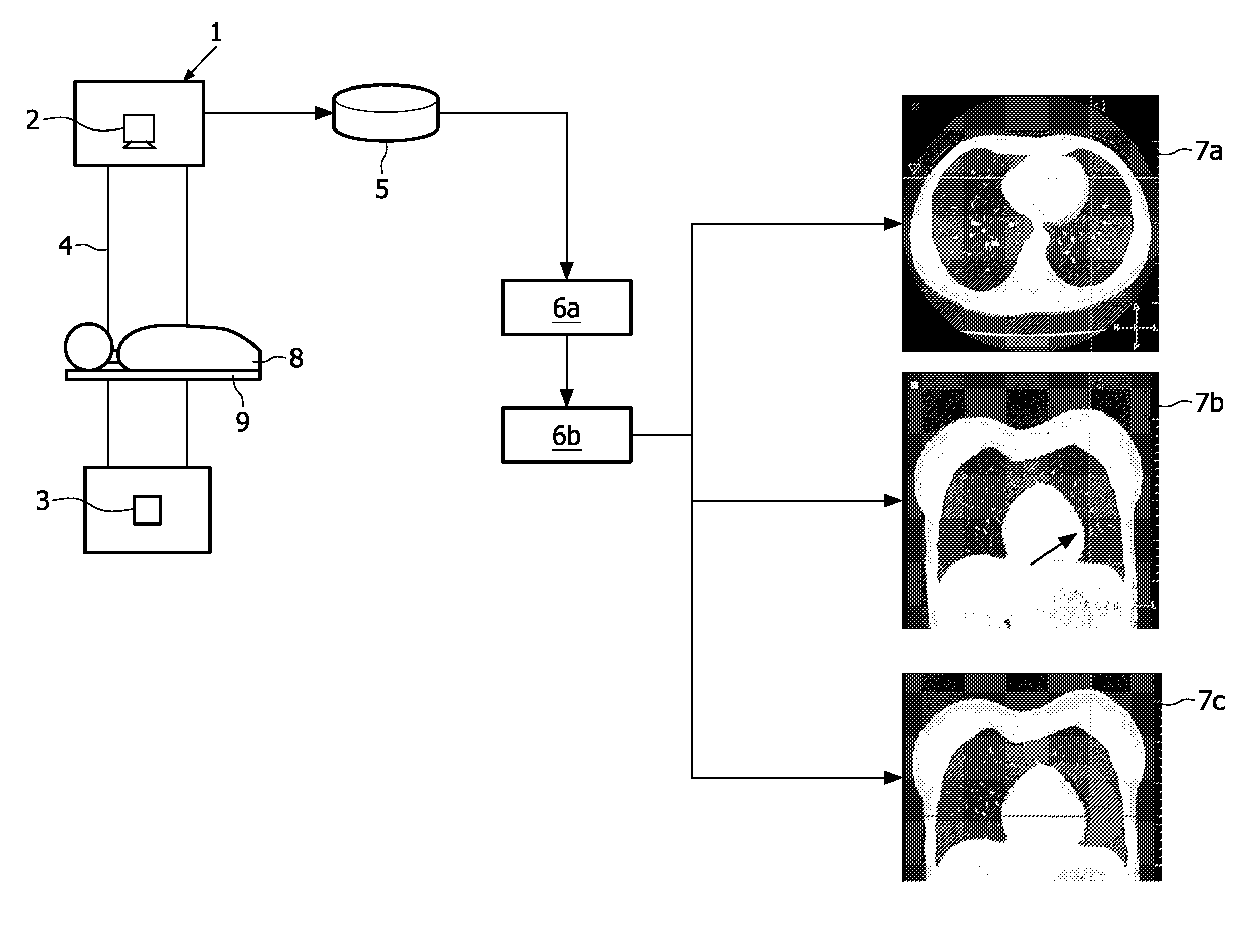
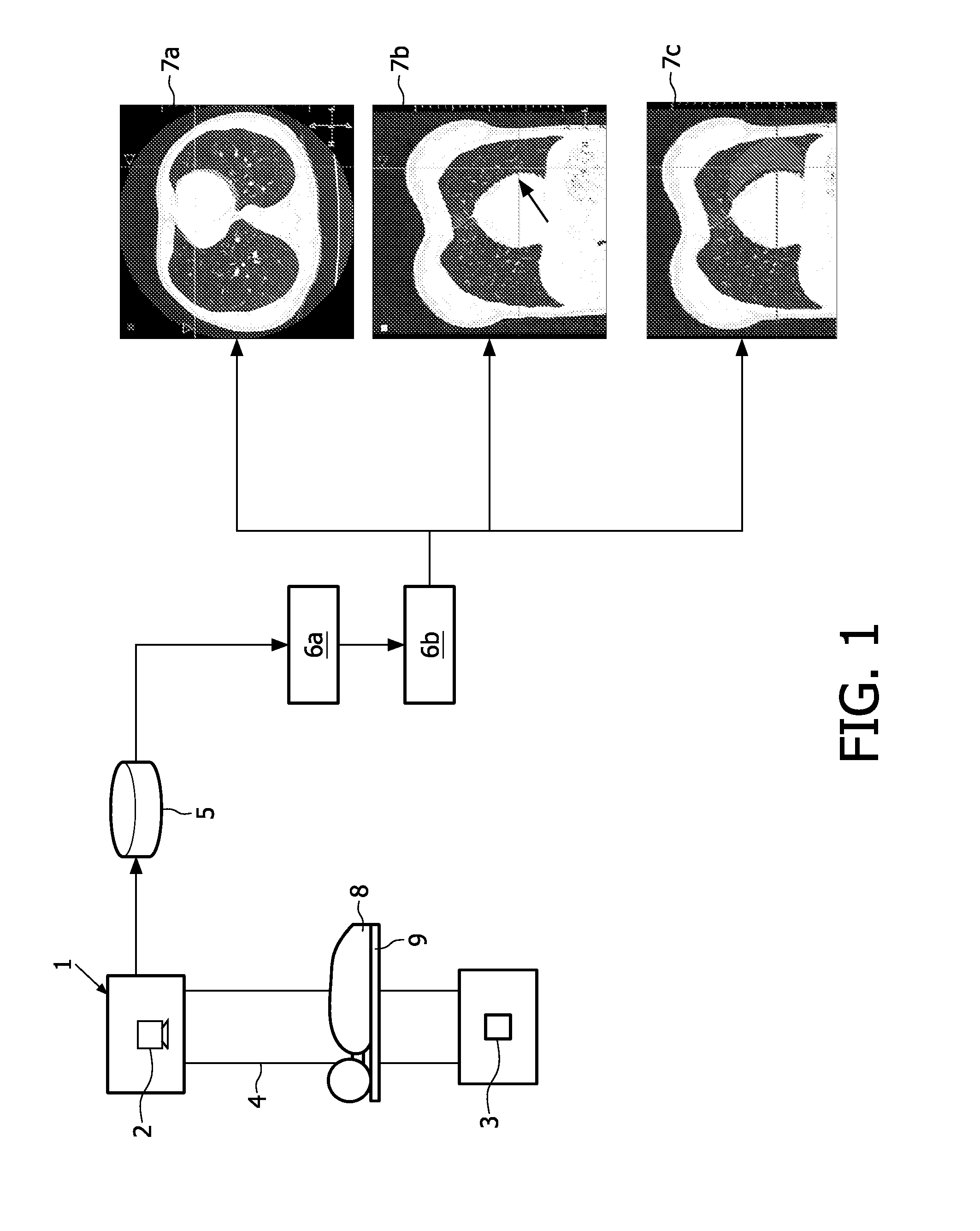
[0030]Referring to FIG. 1, a medical imaging data processing apparatus has a computer tomography (CT) imaging apparatus 1, x-ray sources 2 and detectors 3 arranged in opposed pairs around a circular frame 4. A processor 5 is adapted to execute at least a first computer code 6a and a second computer code 6b producing medical imaging data 7a, 7b and 7c that is displayed on a display device (not shown) in order to assist a physician or radiologist to identify and / or classify features of interest.

[0031]A patient 8 is supported on a platform 9 which can be moved in the longitudinal axis of the platform 9 relative to the frame 4 by means of a motor (not shown) under the control of a control unit (not shown). Data detected by the detectors 3 is input to the processor 5. The processor 5 processes the data received using first computer code 6a and second computer code 6b to provide imaging data output 7a, 7b or 7c. The image data output 7a, 7b or 7c is displayed by a display unit (not shown)...


PUM


Abstract

A data structure for use by a computer system (5) for processing medical image data and representing at least one first region of a patient (8) including at least one computer-detected feature of interest is disclosed. The data structure comprises a first computer code that is executable to detect first data representing at least one second region included within a respective first region. At least one said feature of interest in said second region has a significant likelihood of representing a computer-detected false positive. The second computer code is executable to provide second data for enabling at least one said first region to be displayed on a display device, such that at least one said second region is displayed on the display apparatus differently from part of said first region not containing features of interest having a significant likelihood of representing computer-detected false positives.

Description

FIELD OF THE INVENTION[0001]The present invention relates to computer aided detection systems and methods, and relates particularly, but not exclusively, to an apparatus and method for suppressing and indicating computer-detected features of interest in anatomical regions with a significant likelihood of containing computer-detected false positives.BACKGROUND OF THE INVENTION[0002]The detection and diagnosis of anatomical features of interest such as pulmonary nodules is a standard procedure in radiological practice. The speed and sophistication of current computer-related systems support development of faster and more sophisticated medical imaging systems. The consequential increase in amounts of data generated for processing, and post-processing, has led to the creation of numerous application programs to automatically analyze the medical image date. Thus, various data processing software and systems have been developed in order to assist physicians, clinicians and radiologists, e...

Claims


Application Information

Patent Timeline
IPC IPC(8): G06K9/00
CPCG06T2207/30061G06T7/0012
Inventor WIEMKER, RAFAELOPFER, ROLANDBUELOW, THOMAS
Owner KONINKLJIJKE PHILIPS NV
Why Eureka
Try Eureka