Optical system for a fundus camera

Inactive Publication Date: 2010-01-21
CARL ZEISS MEDITEC AG
View PDF4 Cites 44 Cited by
  • Summary
  • Abstract
  • Description
  • Claims
  • Application Information

AI Technical Summary

Benefits of technology

[0030]The use of the freeform mirror optics in devices with OCT, confocal or line laser scanners or in device and/or methods for imaging the retina can also be advantageous.
[0031]Further, application in confocal laser scanners is advantageous.
[0032]Further, application of the freeform mirror optics in opthalmological devices, particularly fundus cameras, with LEDs operating in continuous operation and/or in pulsed operation as illumination source is advantageous. Aside from eliminating reflections in the recordings, a suppression of motion blur can be successfully carried out because of the short exposure times which can be achieved by the LEDs.
[0033]In particular, by minimizing the distance between the patient's eye being examined and the mirror next to the patient, the overall dimensions of the optical system can also be minimized.
[0034]When using an optical imaging system comprising mirrors, no unwanted reflections occur at optical interfaces. Accordingly, there is no longer any need to block out portions of light in the illumination optics. Unwanted ring

Problems solved by technology

Reflections at the cornea and at the surfaces of the opthalmoscope lens pose a special problem in the observation and recording of the fundus because the light which is reflected by the retina and which carries the information of actual interest has a substantially lower intensity than the light which is reflected before entering the eye.
This type of reflection suppression has come to be known as “anti-reflection point.” A disadvantage of this concept is the proximity of the anti-reflection point to the field diaphragm.
Ring-shaped shadows occur which impair the image impression and, therefore, impede evaluation by the doctor.
This optical system requires a substantial outlay for mechanical mounts for the individual lenses of the objective.
Further, transverse chromatic aber

Method used


Image

  • Optical system for a fundus camera
  • Optical system for a fundus camera
  • Optical system for a fundus camera

Examples


Example

[0039]It is to be understood that the figures and descriptions of the present invention have been simplified to illustrate elements that are relevant for a clear understanding of the present invention, while eliminating, for purposes of clarity, many other elements which are conventional in this art. Those of ordinary skill in the art will recognize that other elements are desirable for implementing the present invention. However, because such elements are well known in the art, and because they do not facilitate a better understanding of the present invention, a discussion of such elements is not provided herein.

[0040]The present invention will now be described in detail on the basis of exemplary embodiments.

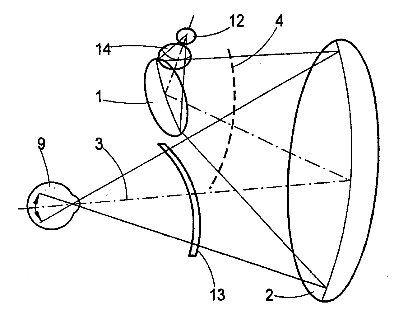
[0041]FIG. 1 is a highly schematic view of a possible advantageous embodiment example of an optical system for implementing reflection-free opthalmoscopy, particularly with a fundus camera. Only the optical elements comprising a substantially coaxial illumination beam path and ...


PUM


Abstract

The invention is directed to an optical system for a fundus camera for reflection-free opthalmoscopy having a beam path with refractive and reflective optical elements which are used substantially in common for illumination and observation or recording. An imaging mirror system substantially comprising a plurality of reflecting optical elements in the form of mirrors and is provided for illuminating and imaging the fundus. At least one optical element, for example, mirror, is formed as a freeform mirror with an imaging, reflecting freeform surface. The optical elements are arranged in a housing in a precisely defined position and attitude relative to one another in such a way that an imaging of the reflecting surfaces of the optical elements on the image of the imaged retina is prevented within a wide diopter range of the patient's eye to be examined.

Description

[0001]The present application claims priority from PCT Patent Application No. PCT / EP2007 / 0011089 filed on Dec. 18, 1007, which claims priority from German Patent Application No. DE 10 2006 061 933.1 filed on Dec. 21, 2006, the disclosure of which is incorporated herein by reference in its entirety.BACKGROUND OF THE INVENTION[0002]1. Field of the Invention[0003]The invention is directed to an optical system for a fundus camera used for the observation and imaging of the ocular fundus. When the ocular fundus is imaged with a camera of this kind, there are generally reflections at the cornea and at surfaces of the imaging optical system which have a negative effect on the quality of the images and the evaluation thereof.[0004]2. Description of Related Art[0005]A fundus camera basically comprises a multi-stage optical system in which an opthalmoscope lens generates an intermediate image that is imaged by a tracking system or main objective onto a film, onto a CCD matrix of a CCD camera,...

Claims


Application Information

Patent Timeline
IPC IPC(8): A61B3/15
CPCG02B17/0621A61B3/156G02B17/086
Inventor KOSCHMIEDER, INGODICK, MANFREDBIERNAT, DETLEFBUCHHEISTER, JANMUELLER, LOTHAR
Owner CARL ZEISS MEDITEC AG
Why Eureka
Try Eureka