Apparatus and method for measuring displacement amount of endoscope image, electronic endoscope, and image processing device for endoscope

a technology of displacement amount and endoscope, which is applied in the field of apparatus and methods for measuring displacement amount of endoscope image, electronic endoscope, and image processing device for endoscope, can solve the problems of endoscope awkward use, endoscope awkward use, and mechanical structures disclosed in the above publications may not be able to cope, so as to reduce the cost of parts used for mounting the solid-state image sensor, the effect of reducing the production cost of electronic endoscope and reducing the cost of reducing

Inactive Publication Date: 2010-02-25
FUJIFILM CORP
View PDF8 Cites 37 Cited by
  • Summary
  • Abstract
  • Description
  • Claims
  • Application Information

AI Technical Summary

Benefits of technology

[0014]It is preferable that the displacement amount obtaining section analyzes the composite image and obtains the displacement amount by automatic measurement. By automatically measuring the displacement amount, burdens of an inspector is reduced, and the displacement amount is measured in a short time.
[0028]Since the mechanical structure is unnecessary, the production costs of the electronic endoscope are reduced. Furthermore, mounting location accuracy of the solid-state image sensor is relaxed. As a result, costs of parts used for mounting the solid-state image sensor are reduced, and production yields of the electronic endoscope are increased.

Problems solved by technology

As a result, the medical instrument projected from the medical instrument outlet is not displayed in a normal position on the monitor, making the endoscope awkward to use.
As a result, the zoomed area is displaced from the intended area, which also makes the endoscope awkward to use.
When the number of pixels in the image sensor further increases, the mechanical structures disclosed in the above publications may not be able to cope with the improvement on zooming alignment required due to the higher magnification of the electronic zoom, and improvement on the mounting location accuracy required due to downsizing of the image sensor in an electronic endoscope with a smaller diameter.
In addition, in the above publications No. 10-028233, 11-290269, and 2001-086378, the mechanical structures increase production costs.

Method used


Image

  • Apparatus and method for measuring displacement amount of endoscope image, electronic endoscope, and image processing device for endoscope
  • Apparatus and method for measuring displacement amount of endoscope image, electronic endoscope, and image processing device for endoscope
  • Apparatus and method for measuring displacement amount of endoscope image, electronic endoscope, and image processing device for endoscope

Examples


first embodiment

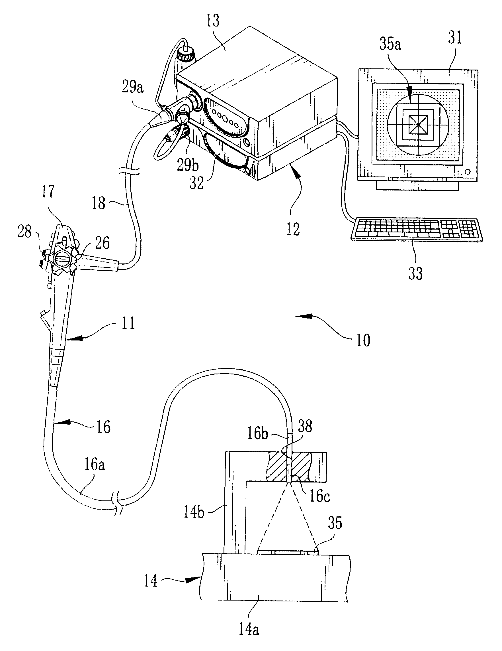
[0127]Next, the tilt angle θ is input through the keyboard 33. Thereby, all of the displacement amount information (the horizontal displacement amount A, the vertical displacement amount B, and the tilt angle θ) is input and stored in the EEPROM 44 of the electronic endoscope 11. The displacement inspection is thus completed. The processing from then on is the same as that in the first embodiment, and the description thereof is omitted.

[0128]As described above, in the displacement inspection of the second embodiment, the measurement of the positional displacement amount and the positional displacement correction are performed first. Accordingly, the tilt angle θ (the rotational displacement amount) of the orientation detection mark 37a is calculated using the mathematical expression (2) that is easier than the mathematical expression (1) of the first embodiment. Thus, the tilt angle θ is calculated easily.

[0129]Next, a third embodiment of the present invention is described. In the f...

third embodiment

[0142]The white area 93a shows a positional displacement of the test chart image 35b in the vertical direction, and is obtained by counting the number of pixels in a width Yp in the vertical direction of the white area 93a. The white area 93b shows positional displacement of the test chart image 35b in the horizontal direction, and is obtained by counting the number of pixels in a width Xp in the horizontal direction of the white area 93b. The counting of the numbers of pixels in the widths XP and YP is performed through the image analysis of the test mask composite image 92 by the displacement amount measuring section 82 as described in the

[0143]When the test chart image 35b is positionally displaced, for example, toward the lower right direction, the upper left direction, the lower left direction, the upper direction, or the like, from the reference position, only the positions and the number of the white areas change, and therefore the descriptions thereof are omitted. The image ...

second embodiment

[0145]When the test chart image 35b is positionally and rotationally displaced from the reference position and the reference orientation, it is necessary to correct the rotational displacement correction prior to measurement of the positional displacement amount (the numbers of pixels in the width Xp and the width Yp). For this reason, unlike the second embodiment, the measurement and correction of the rotational displacement are performed first.

[0146]In the third embodiment, the test chart image and the test mask image other than those illustrated in FIGS. 13A to 13D, for example, the test chart image 35c and the test mask image 99 shown in FIGS. 14A and 14B can be used. An endoscope image 30c displays an observation image 94 containing a test chart image 35c. The test chart image 35c consists of a red dot mark 95 indicating the center C1 of the test chart image 35c and a green dot mark 97 indicating orientation of the test chart image 35c. Hereinafter, the red dot mark 95 may be r...


PUM


Abstract

An image of a test chart having a test pattern is taken with an electronic endoscope. A DSP of a processing device generates a test pattern image from image signals input from the electronic endoscope. An image compositor composites the test pattern image and a test mask image having a predetermined reference pattern and generates a test mask composite image. An inspector visually measures positional and rotational displacement amounts of the test pattern with respect to the reference pattern in the test mask composite image displayed on a monitor, and inputs measurement results as displacement amount information to the processing device. The displacement amount information is stored in an EEPROM of the electronic endoscope. The processing device calibrates an endoscope image of a body cavity based on the displacement amount information input from the electronic endoscope.

Description

FIELD OF THE INVENTION[0001]The present invention relates to an electronic endoscope, an image processing device for performing image processing to an endoscope image obtained with this electronic endoscope, and an apparatus and a method for measuring a displacement amount of the endoscope image.BACKGROUND OF THE INVENTION[0002]Electronic endoscope systems are widely used in medical diagnoses. An electronic endoscope system is composed of an electronic endoscope having an insert section to be inserted in a body cavity of a patient and a processing device connected to this electronic endoscope. The processing device generates an endoscope image based on image signals received from an image sensor mounted to a distal portion of the insert section of the electronic endoscope. The processing device performs image processing to the endoscope image, and then outputs the endoscope image to a monitor. A doctor observes the body cavity and makes diagnosis, or treats the patient using a medic...

Claims


Application Information

Patent Timeline
Patent Type & Authority Applications(United States)
IPC IPC(8): A61B1/04
CPCA61B1/00057A61B1/05A61B1/04
Inventor SHIDARA, KENICHI
Owner FUJIFILM CORP
Why Eureka
Try Eureka