Reconstituted mineralized cartilage tissue

a mineralized cartilage and tissue technology, applied in the field of reconstituted mineralized cartilage tissue, can solve the problems of hampered study of the calcified cartilage zone, lack of in vitro culture system, and inability to study the tissue formed by these cells, and achieve the effect of enhancing the healing of a bone fracture in a patien

Inactive Publication Date: 2010-03-18
MOUNT SINAI HOSPITAL
View PDF2 Cites 3 Cited by
  • Summary
  • Abstract
  • Description
  • Claims
  • Application Information

AI Technical Summary

Benefits of technology

[0019]The invention also contemplates a method of replacing or repairing damaged or deficient cartilage in a joint of a patient comprising implanting a mineralized biological material or reconstituted mineralized cartilaginous tissue of the invention in the joint of the patient. Methods for enhancing healing of a bone fracture in a patient are contemplated which comprise inserting a mineralized biological material or reconstituted mineralized cartilaginous tissue of the invention into the site of a fracture.

Problems solved by technology

Study of the calcified cartilage zone has been hampered, in part, by the lack of an in vitro culture system.
The tissue formed by these cells is not well suited to the study of articular cartilage mineralization as the cartilage serves as a template for bone formation.
Therefore, it is not appropriate to extrapolate observations generated from these mineralizing cultures to the calcified articular cartilage.

Method used


Image

  • Reconstituted mineralized cartilage tissue
  • Reconstituted mineralized cartilage tissue
  • Reconstituted mineralized cartilage tissue

Examples


example 1

Characterization of the Mineralized Chondrocyte Cultures

[0067]Determination of the Enrichment of Chondrocytes from the Deep Zone: As alkaline phosphatase activity is detected in cells at or above the calcified zone of articular cartilage, this enzyme was used as a marker to assess the enrichment of these cultures with cells from the deep cartilage. Cells with an alkaline phosphatase activity of at least 2 μM PNP / hr / 106 cells, which represented at least a fourfold increase in alkaline phosphatase activity when compared to the alkaline phosphatase activity detected in the same number of cells obtained from the entire cartilage (data not shown), were used to establish the cultures. Cells with alkaline phosphatase activity less than this did not mineralize as well or as rapidly. On average the alkaline phosphatase activity of the cells isolated from the deep layer of cartilage was 4.1±0.8 μM PNP / hr / μg DNA (mean±SE). In contrast the alkaline phosphatase activity of cells isolated from th...

example 2

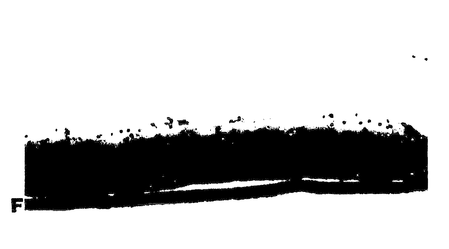
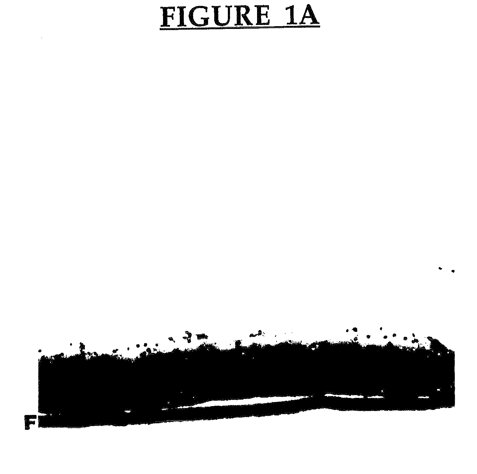
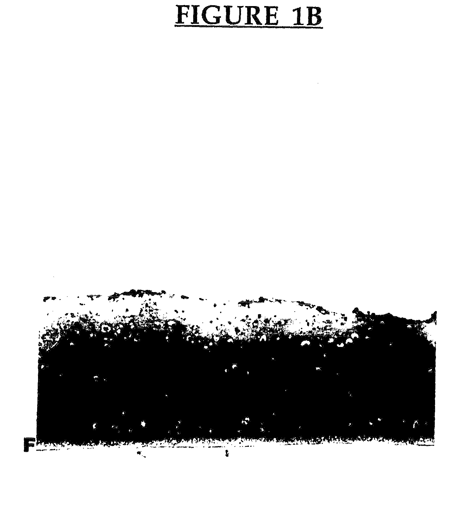
[0082]Chondrocytes from the mid and superficial zones of articular cartilage were cultured on top of the mineralized cartilaginous tissue described above. Chondrocyte cultures are grown in Hams F12 medium containing 20% fetal bovine serum as described above, and after about five days the medium is changed to DMEM with 20% fetal bovine serum supplemented with 10 mM β-glycerophosphate, 100 μg / ml ascorbic acid, and 25 mM Hepes buffer. The cultures are then maintained for six weeks or longer. This results in a reconstituted mineralized cartilage tissue which has a deep mineralized layer and mid and superficial non-mineralized layers substantially similar to articular cartilage tissue in vivo. In particular, FIG. 5 is a photomicrograph of 49 day old culture showing superficial and midzone chondrocytes cultured on cartilaginous tissue formed by the deep cells. Cartilaginous tissue is present and mineral deposits are present in the tissue generated by the deep cells only (Von Kossa stain)....


PUM

PropertyMeasurementUnit
pore sizeaaaaaaaaaa
concentrationaaaaaaaaaa
timeaaaaaaaaaa

Abstract

A biological material comprising a continuous layer of cartilaginous tissue reconstituted in vitro which contains components associated with cartilage mineralization. The biological material may be cultured with a mineralizing agent to form a mineralized biological material. The mineralized biological material is characterized by having a biochemical composition and physiological organization substantially similar to the deep and contiguous calcified cartilage zones of articular cartilage found in animals in vivo. Methods for preparing the biological materials and methods of using the biological materials are described.

Description

CROSS-REFERENCE TO RELATED APPLICATIONS[0001]This application is a continuation of U.S. patent application Ser. No. 10 / 121,738, filed Apr. 12, 2002, which is a continuation of U.S. patent application Ser. No. 09 / 068,233, filed Sep. 29, 1998, now U.S. Pat. No. 6,464,729, issued Oct. 15, 2002, which is the US national stage of International Patent Application No. PCT / CA96 / 00729, filed Nov. 5, 1996. International Patent Application No. PCT / CA96 / 00729 is a continuation-in-part of U.S. patent application Ser. No. 08 / 706,625, filed Sep. 6, 1996 and claims the benefit of the priority of U.S. Provisional Patent Application No. 60 / 007,279, filed Nov. 6, 1995, now expired. The priority applications described above are herein incorporated by reference.BACKGROUND OF THE INVENTION[0002]Articular cartilage is a specialized tissue found at the end of articulating bones. It is responsible for the distribution of load resistance to compressive forces, and the smooth gliding that is part of joint fun...

Claims


Application Information

Patent Timeline
Patent Type & Authority Applications(United States)
IPC IPC(8): A61K9/00A61P19/02A61K35/12A61K48/00A61L27/38C12N5/00C12N5/077
CPCA61K35/12C12N2533/90A61L27/3608A61L27/3633A61L27/365A61L27/3654A61L27/3817A61L27/3847A61L27/3852A61L27/3895A61L2430/06C12N5/0068C12N5/0655C12N2500/42A61K48/00A61P19/02
Inventor KANDEL, RITA
Owner MOUNT SINAI HOSPITAL
Why Eureka
Try Eureka