Method for diagnosis of a disease involving an Anti-at1-receptor antibody

a technology of at1 receptor and antibody, applied in the field of method for diagnosis of a disease involving an at1 receptor antibody, can solve the problems of severe complications of rheumatoid arthritis and difficulty in standard treatment methods

Active Publication Date: 2010-04-22
CELLTREND
View PDF7 Cites 7 Cited by
  • Summary
  • Abstract
  • Description
  • Claims
  • Application Information

AI Technical Summary

Benefits of technology

[0017]In a preferred embodiment of the invention the determination of presence or absence of an anti-AT1-receptor antibody is done by detecting one or mo

Problems solved by technology

All patients with rheumatic diseases displayed severe compl

Method used


Image

  • Method for diagnosis of a disease involving an Anti-at1-receptor antibody
  • Method for diagnosis of a disease involving an Anti-at1-receptor antibody
  • Method for diagnosis of a disease involving an Anti-at1-receptor antibody

Examples


example i

AT1-Receptor ELISA

[0084]A suitable streptavidine-coated microtiter plate is loaded with the biotinylated peptide encoding the AT1-receptor.

[0085]Such peptides may be AVHYQSN (SEQ ID NO. 1), SHFYQTR (SEQ ID NO. 2) and / or GYYFDTN (SEQ ID NO. 3).

[0086]To this end, 100 μl of a solution per well in the microtiter plate is incubated with 5 μg / ml in a suitable dilution buffer. To measure the non-specific binding, wells are also filled with 100 μl of dilution buffer. The dilution buffer may comprise 0.5% bovine serum albumin, 10 mM phosphate buffer (pH 7.4), 140 mM NaCl and 0.05% TWEEN 20.

[0087]Subsequent to the time period of reaction, the peptide solution is moved by decanting and each well is washed three times with 250 μl of a suitable wash buffer. A suitable wash buffer may comprise 10 mM phosphate buffer (pH 7.4), 140 mM NaCl and 0.1% TWEEN 20. Thereafter, 100 μl per well of sera are diluted in a dilution buffer and placed on both the peptide-loaded and comparative plates are incubate...

example ii

[0090]Various patient groups (with positive diagnosis) as well as controls were tested with respect to the presence or absence of the anti-AT1-receptor antibody. Table I shows the result obtained.

TABLE IPresence of anti-AT1-receptorDiseaseantibody in %rheumatic disease35Patients without inflammatory rheumatic8.3disease

example iii

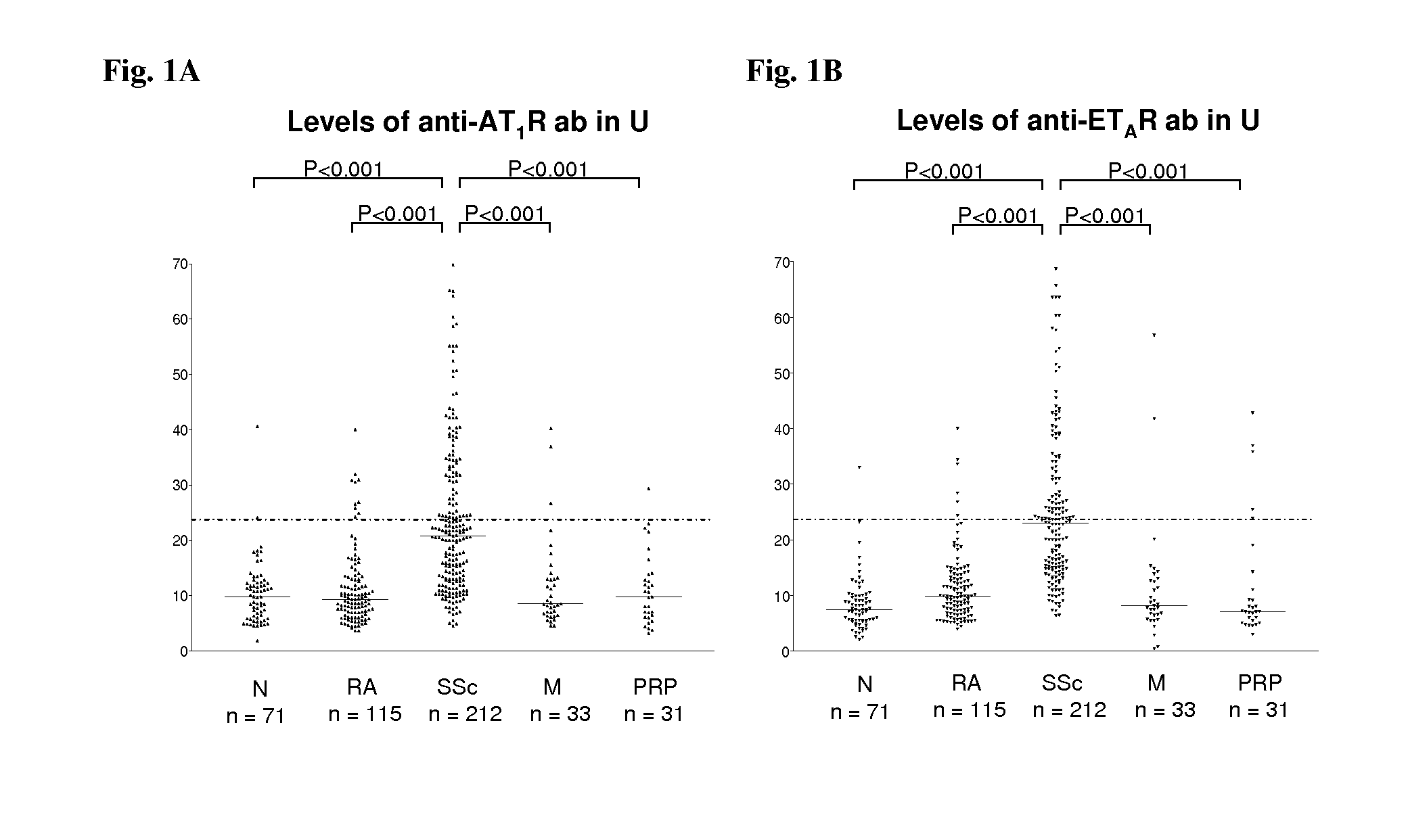
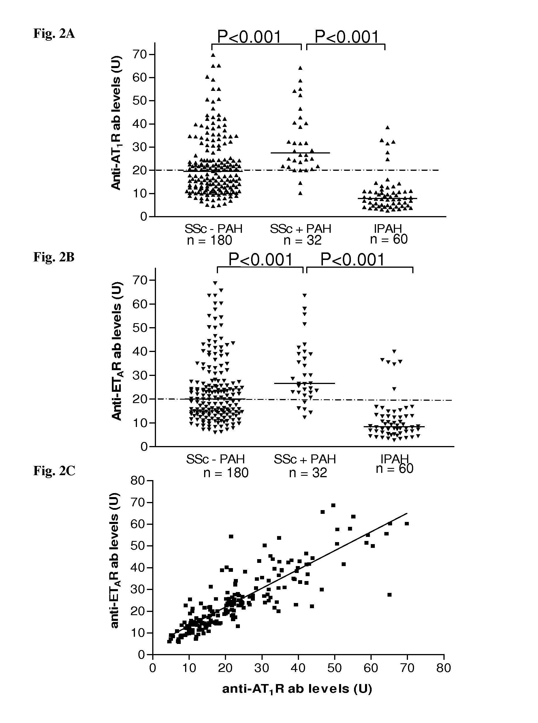
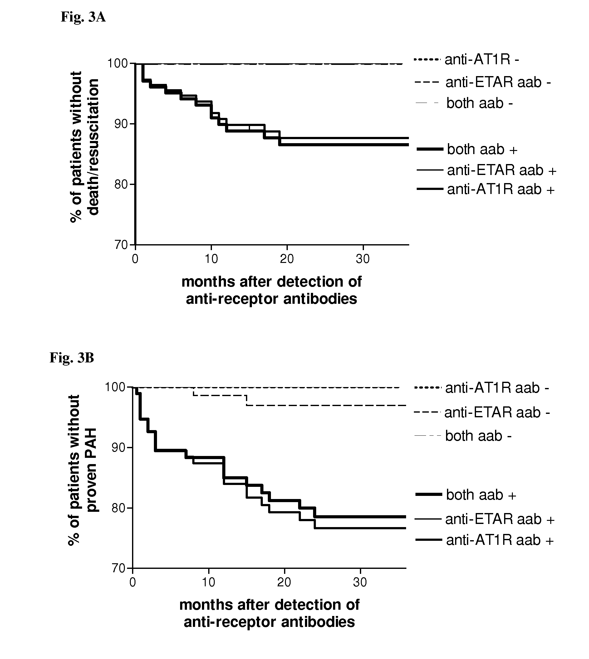
[0091]We analyzed sera of 212 patients with systemic sclerosis (SSc), 60 healthy control subjects, 120 rheumatoid arthritis patients and 124 additional control subjects with either morphea, primary Raynaud phenomenon or idiopathic pulmonary hypertension for the presence of anti-bodies directed against angiotensin II type 1 receptor (AT1R) and endothelin-1 type A receptor (ETAR) by a solid phase assay. Individual organ involvement, patient survival and the effect of immunosuppressive or cell therapies were also assessed. Vascular responsiveness to natural receptor ligands, Angiotensin II and Endothelin-1 was studied ex vivo in pulmonary resistance arteries.

[0092]AT1R-AA was detected in 56.6% of the SSc patients, respectively, but only in 9.6% subjects with control diseases, and 2.8% of the normal subjects. Patients with AT1R-AA had more severe disease. Positivity for AT1R-AA strongly predicted development of pulmonary arterial hypertension and SSc-related mortality. The antibody leve...


PUM


Abstract

The present invention relates to a method for diagnosis of a disease selected from the group of diabetes, vasculitis, collagenosis, an inflammatory rheumatic disease and arteriosclerosis wherein, presence or absence of an anti-AT1-receptor antibody is determined in a sample from a patient to be diagnosed and wherein, the presence of an anti-AT1-receptor antibody is indicative of the disease. The invention further relates to the use of an inhibitor of an anti-AT1-receptor antibody or an inhibitor of an AT1-receptor for the production of a medicament as well as plasmapheresis of blood for the removal of anti-AT1-receptor antibodies.

Description

[0001]In medicine, vasculitis (plural: vasculitides) is a group of diseases featuring inflammation of the wall of blood vessels due to leukocyte migration and resulting damage. While most vasculitides are rare, they generally effect several organ systems and can cause severe disability. Vasculitides are classified into the large vessel vasculitis such as Takayasu arteritis and giant cell (temporal) arteritis. Further, medium vessel vasculitis, such as polyarteritis nodosa and Kawasaki disease. Small vessel vasculitis such as Churg-Strauss arteritis, Wegener's granulomatosis and hypersensitivity vasculitis.[0002]Patients usually present with systemic symptoms with single or multi organ dysfunction. Common (and nonspecific) complaints include fatigue, weakness, fever, arthralgias, abdominal pain, hypertension, renal insufficiency, a neurologic dysfunction. The following symptoms should raise a strong suspicion of vasculitis, however, the diagnosis is difficult. These symptoms are mono...

Claims


Application Information

Patent Timeline
IPC IPC(8): A61K39/395G01N33/53C07K2/00A61K31/4178A61K31/4184A61K31/41A61K31/7088A61P37/00
CPCG01N33/564G01N2800/10G01N2333/72A61P37/00
Inventor SCHULZE-FORSTER, KAIHEIDECKE, HARALD
Owner CELLTREND
Why Eureka
Try Eureka