

Differential diagnosis for scleroderma
a differential diagnosis and scleroderma technology, applied in the field of scleroderma differential diagnosis, can solve the problem that no data are available on the presence of these autoantibodies
- Summary
- Abstract
- Description
- Claims
- Application Information
AI Technical Summary
Benefits of technology
Problems solved by technology
Method used
Image

Examples
example 1
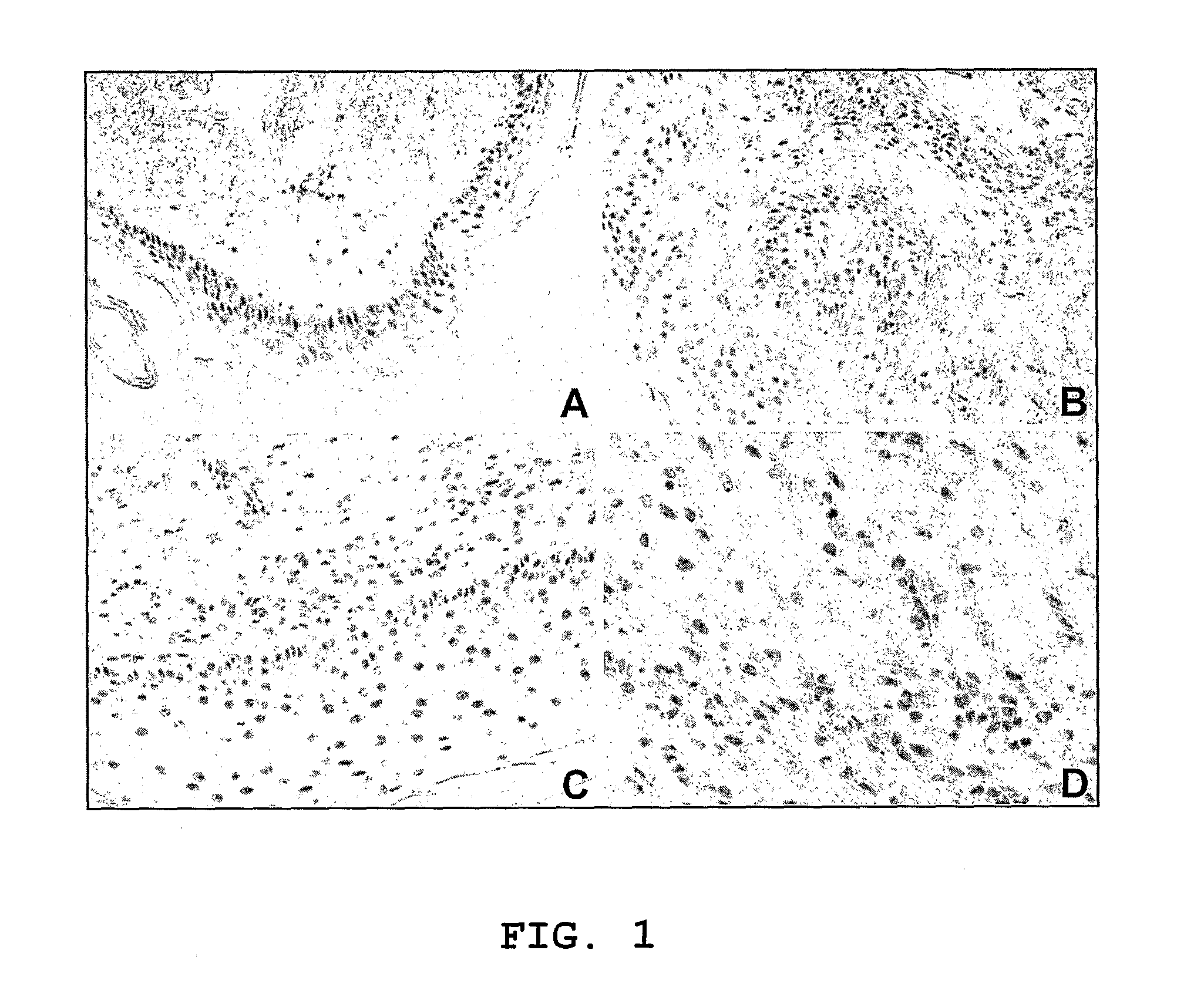
Anti-IFI16 Autoantibody Levels by ELISA and Skin IFI16 Immunoreactivity were Elevated in SSc and SLE
[0043]As recently reported in the normal epidermis from healthy controls, IFI16 expression was restricted to the basal layer (FIG. 1A). Notably, as shown in the representative sections in FIG. 1, IFI16 expression was greatly increased and found ubiquitously expressed in all layers of the epidermis in the lesional skin from both SSc (FIG. 1B) and SLE (FIG. 1C) patients. Furthermore, the dermal inflammatory infiltrate showed IFI16 positive staining, indicating that it is expressed to a high level in lymphocytes, fibroblasts and endothelial cells.
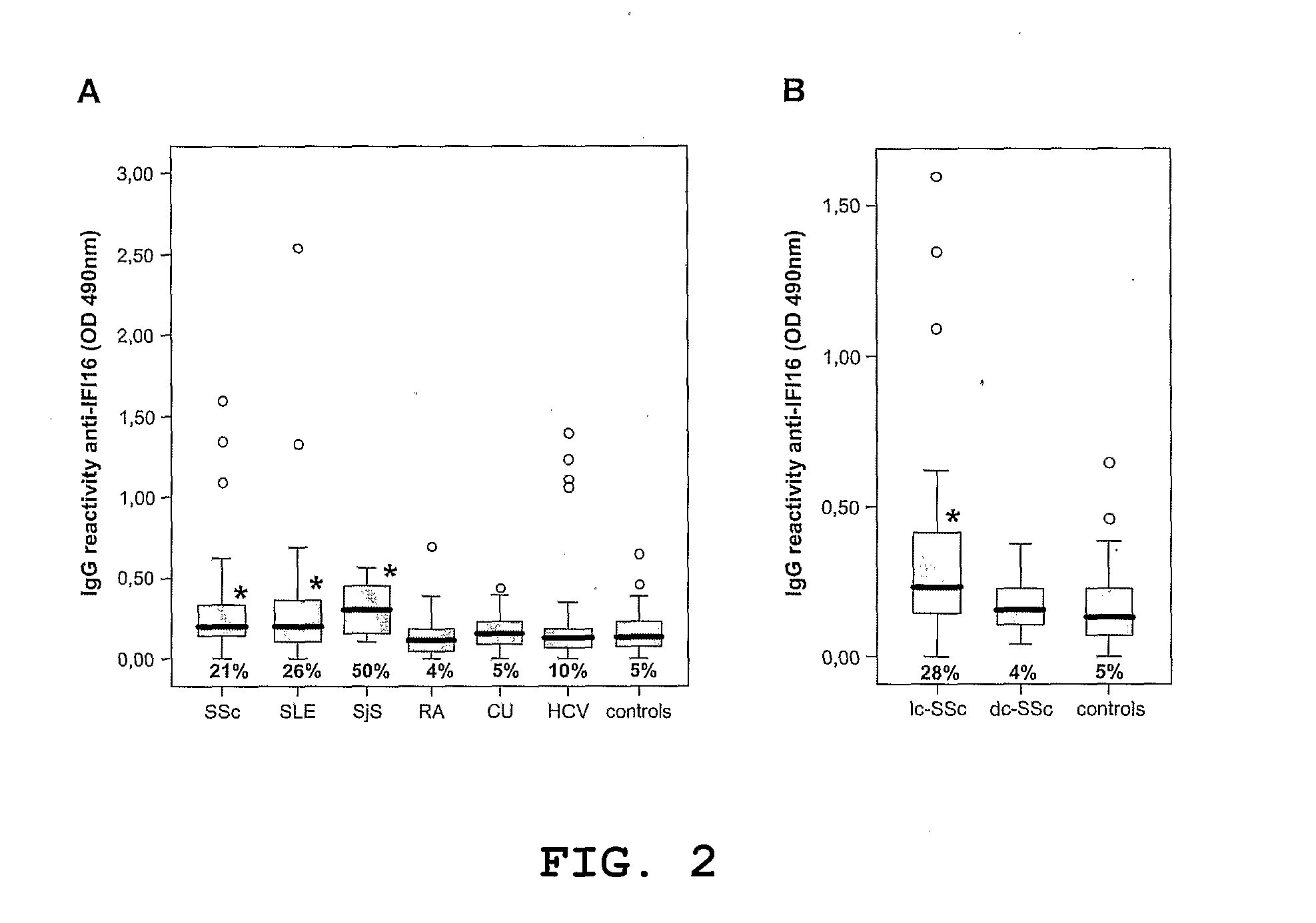
[0044]To verify whether the increased expression of IFI16 in the affected skin was related to the presence of anti-IFI16 autoantibodies, their presence and levels in serum samples from 100 SLE patients and 82 SSc patients were assessed using ELISA with recombinant IFI16 protein. Other autoimmune diseases, including SjS, RA and CU as well as heal...
example 2
Association of anti-IFI16 Antibodies with Clinical Parameters
[0045]Since the presence of anti-IFI16 autoantibodies had already been reported in both SjS and SLE, where a significant serological heterogeneity is well known to occur (14), but not in SSc, a decision was taken to gain more insight into the actual role of anti-IFI16 autoantibodies in SSc pathogenesis. Univariate analysis showed that anti-IFI16 autoreactivity in SSc patients was not associated with either disease duration or disease severity as measured by Medsger stage, HAQ disability index, organ involvement and positivity to other autoantibodies (data not shown). By contrast, a strict association between anti-IFI16 reactivity and the cutaneous form of the disease was found, with patients in the limited cutaneous scleroderma (lc-SSc) having higher anti-IFI16 IgG titers than patients with the diffuse form (dc-SSc) (p=0.017). Indeed, as shown in FIG. 2B, anti-IFI16 titers above the 95th percentile of the controls were obs...
example 3
Logistic Regression Model and Sensitivity-Specificity Analysis for lc-SSc Prediction
[0047]In our samples the cutaneous form of SSc was significantly associated with both anti-centromere (χ2=18.771 p2=32.689 p<0.0005) autoantibodies. Logistic regression showed that all the three serological markers were independent predictors of the cutaneous form of scleroderma, and their combination was able to explain 62% of the associated variability. This model was able to correctly predict 89% of the clinical presentation forms of scleroderma. Moreover, anti-IFI16 reactivity displayed lower sensitivity (28%) and higher specificity (96%) than those found with either anti-topoisomerase I (95% and 67% respectively) or anti-centromere (65% and 92% respectively). The combined use of anti-IFI16 and anti-centromere markers gave rise to the highest sensitivity and specificity score (79% and 92% respectively). Interestingly, in the SSc subgroup negative for both anti-centromere and anti-topoisomerase I ...
PUM
 Abstract
Description
Claims
Application Information