Medical implantable lead and a method for attaching the same

Active Publication Date: 2010-05-06
ST JUDE MEDICAL
View PDF7 Cites 29 Cited by
  • Summary
  • Abstract
  • Description
  • Claims
  • Application Information

AI Technical Summary

Benefits of technology

[0013]It is an object of the present invention to provide a fixation arrangement for a medical implantable lead, having a helix, which can be manufactured of a thin

Problems solved by technology

One disadvantage with a fixation device like this is that the impedance between each helix and the tissue will be small.
A small impedance will lead to increased energy consumption and hence more frequent replacement of the implanted electronic devices due to discharged batteries.
In some applications measures may have to be taken to avoid this problem, such as partly insulating of the helices as is disclosed in the document, which will lead to increased costs for manufacturing.
This trend has resulted in side-effects, s

Method used


Image

  • Medical implantable lead and a method for attaching the same
  • Medical implantable lead and a method for attaching the same
  • Medical implantable lead and a method for attaching the same

Examples


first embodiment

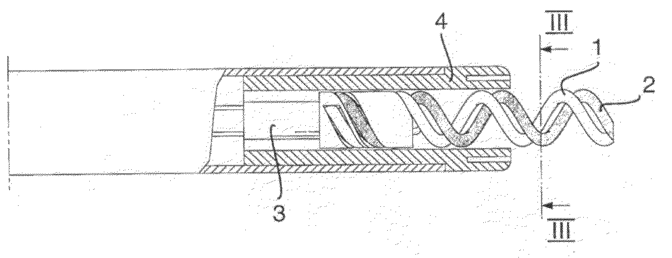
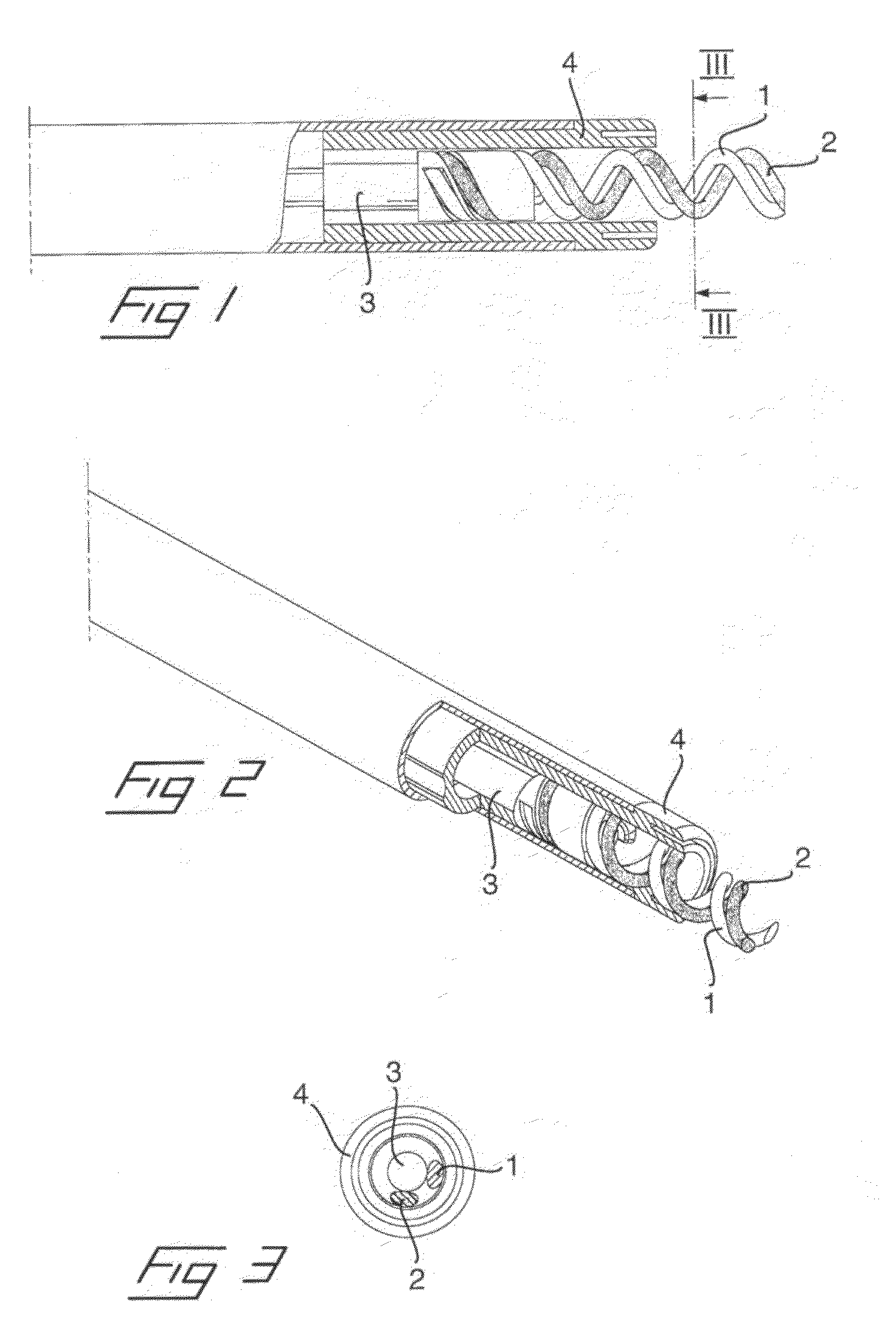
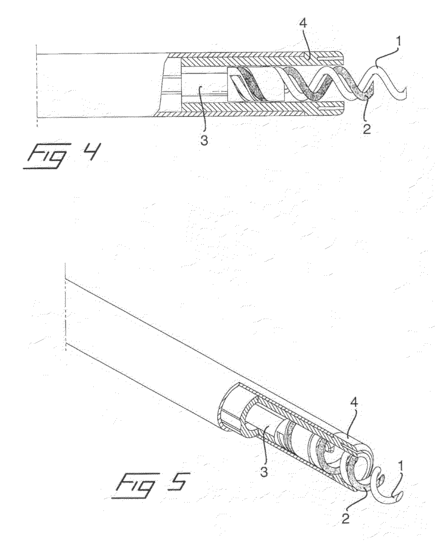
[0029]an arrangement according to the invention is shown in FIGS. 1-3. In accordance with the present invention, a dual helix fixation arrangement has two fixation helices 1, 2, each formed of a helical wire and each terminating in a sharp point that is capable of penetrating the endocardium and the myocardium of a heart.

[0030]The two helices are mounted in a holder portion on a shaft 3 that can be rotated from the opposite, proximal end of the lead or the implantation device by means of for example a rotatable coil or a stylet in a way commonly known in the art. When the shaft 3 and the two helices 1, 2 are rotated, this causes them to advance from inside a protective covering or header 4, in which they are initially positioned, so as to penetrate into the endocardium and the myocardium. In the illustrated embodiment the two helices have the same diameter and pitch but are displaced by about 90° in relation to each other, as is best seen in FIG. 3, in order to position the helical ...

third embodiment

[0032]In FIGS. 6 and 7 is disclosed the invention. Here the first and second helices 1, 2 have the same length and are displaced 180° in relation to each other. However, in order to increase the impedance of the second helix 2, it has been manufactured of a thinner wire than the first helix 1. Accordingly, the first helix 1 can be formed of a helical wire of a desirable cross-sectional dimension to achieve an optimal fixation to the tissue, whereas the second helix 2 can be formed of a helical wire of a desirable cross-sectional dimension to achieve an optimal electrical connection to the tissue with respect to e.g. impedance and capture threshold.

[0033]Since the dual helices of all three of the embodiments described above are mounted with a distance in relation to each other, over-rotating of the header / helix combination is prevented when torque is applied to the helix during implantation. This is due to the fact that by using dual helices with a proper pitch, two entry punctures a...


PUM


Abstract

A medical implantable lead of the type being adapted to be implanted into a human or animal body and attached with a distal end to an organ inside the body, has a helix of a helical wire in the distal end which is adapted to be screwed into the organ. In addition to the first helix, the lead also has a second helix of a helical wire, the second helix having the same diameter, the same pitch and being intertwined with the helical wire of the first helix and which, upon rotation of the first helix, will be rotated and screwed into the tissue. The first helix is electrically non-conductive whereas the second helix is electrically conductive. In a method for attaching a medical implantable lead to an organ inside a human or animal body, such a medical lead is employed and fixed to tissue in vivo.

Description

BACKGROUND OF THE INVENTION[0001]1. Field of the Invention[0002]The invention relates to a medical implantable lead of the type being adapted to be implanted into a human or animal body and attached with a distal end to an organ inside the body, and having a helix of a helical wire in the distal end which is adapted to be screwed into the organ, wherein the lead, in addition to the first helix, also comprises a second helix of a helical wire, the second helix having the same pitch and being intertwined with the helical wire of the first helix and which, upon rotation of the first helix, will be rotated and screwed into the tissue.[0003]The invention also relates to a method for attaching a medical implantable lead to an organ inside a human or animal body, by screwing a helix of a helical wire into the tissue of the organ.[0004]The invention is primarily adapted for attaching the tip of a cardiac electrode into cardiac tissue for connecting e.g. a pacemaker or defibrillator to a hea...

Claims


Application Information

Patent Timeline
IPC IPC(8): A61N1/05
CPCA61N1/0573
Inventor HILL, ROLFSTEGFELDT, OLOF
Owner ST JUDE MEDICAL
Why Eureka
Try Eureka