Calibrated rpma assay

a colorimetric system and rpma technology, applied in material analysis, instruments, measurement devices, etc., can solve problems such as poor colorimetric system dynamic rang

Inactive Publication Date: 2010-08-12
GEORGE MASON INTPROP INC
View PDF0 Cites 4 Cited by
  • Summary
  • Abstract
  • Description
  • Claims
  • Application Information

AI Technical Summary

Problems solved by technology

However, because of the very poor dynamic range of colorimetric systems, the ability to determine the concentration of an analyte in an input sample so that antigen-antibody interactions are within the linear dynamic range requires that the sample be printed in a miniature dilution curve, usually a series of 4 to 5 1:2 dilutions.

Method used


Image

  • Calibrated rpma assay
  • Calibrated rpma assay

Examples


example i

Materials and Methods

[0096]Methods for preparing, assaying, and analyzing samples for a method of the invention are conventional and well-known to skilled workers. Some of these methods are described below:

Laser Capture Microdissection (of tumor cells): 8 um frozen sections are prepared on either glass or membrane slides. Frozen sections are fixed in 70% ethanol, stained with Mayer's Hematoxylin and Scott's Tap Water Substitute, and dehydrated in gradient ethanol, with a final clearing in xylene.

[0097]The slides are rapidly air dried and tumor cells are isolated by laser capture microdissection (Pixcell™ and Veritas™, Arcturus Molecular Devices; CA, USA). Formalin or alcohol fixed paraffin embedded specimens can also be used, and subjected to Laser Capture Microdissection as above with appropriate tissue stains.

Reverse Phase Protein Microarrays. Microdissected cells are subjected to lysis in boiling 2.5% beta-mercaptoethanol in T-PER (Pierce, Rockford, Ill.) mixed 1:1 with 2×SDS Tri...

example ii

An Illustrative Assay of 26 Analytes

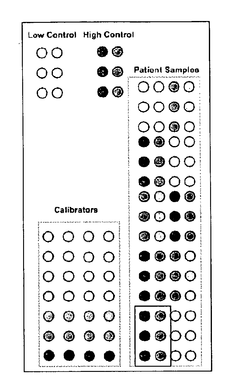
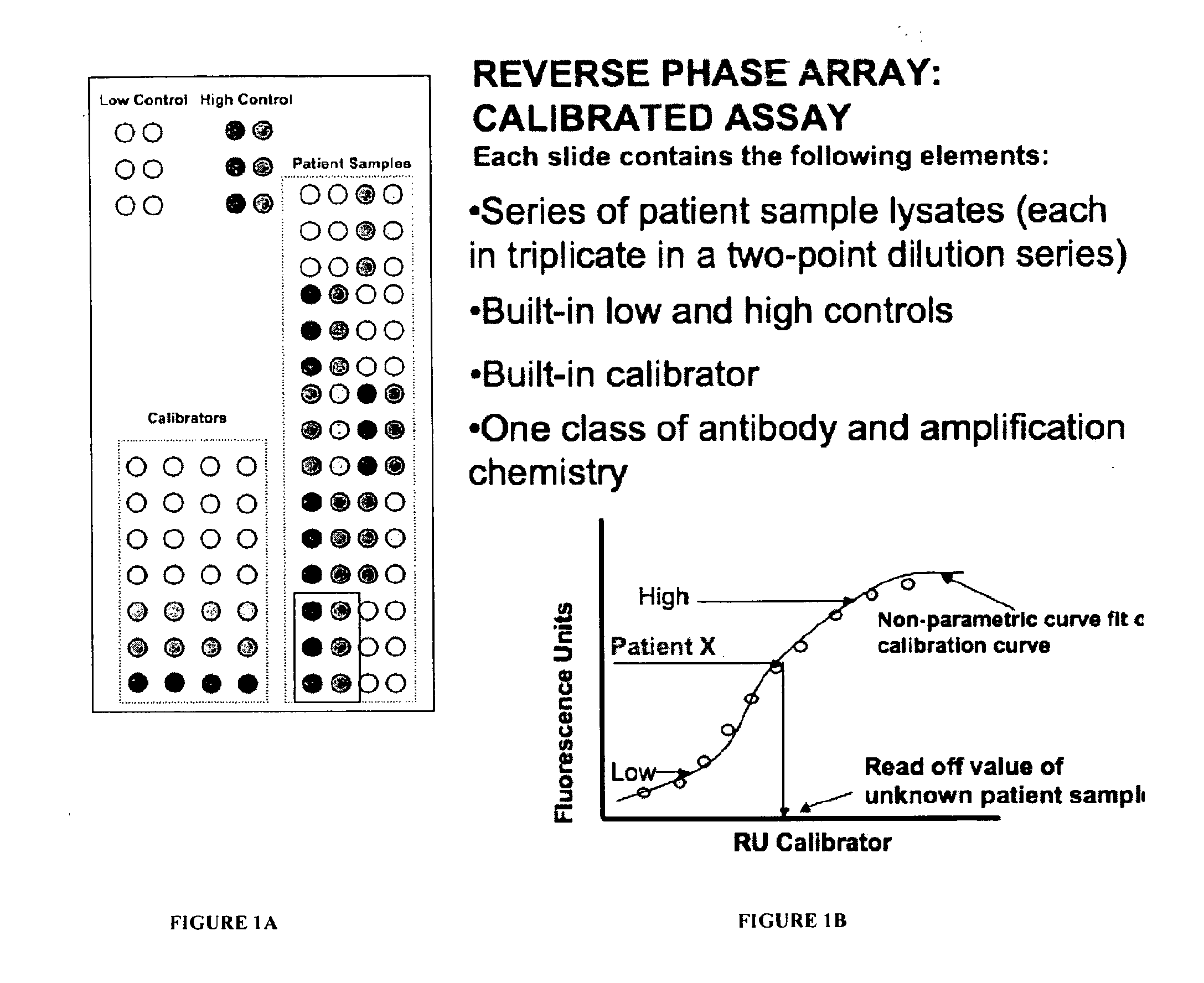
[0098]In the present Example, tissue samples from 2 to about 500 patients are prepared and lysed, and aliquots of the whole cell lysates are printed onto slides in a microarray, as described in Example I. Each lysate is printed as a neat and as a 1:4 dilution, each in triplicate. The lysates are printed onto each of 27 slides, each one for the determination of a different analyte in the sample, as well as one slide that is stained for total protein (e.g Sypro Ruby Blot Stain, Molecular Probes Eugene, Oreg.). The analytes to be analyzed / quantitated are:

1. Total EGFR

[0099]2. Total c-erbB2

3. Total VEGFR2 (KDR, Flk-2)

4. Total PDGFRbeta

5. Total PDGFRalpha

6. Total FLT3

7. Phospho FLT-3 (Y5899 / Y591)

8. Phospho VEGFR2 (Y1212)

9. Phospho VEGFR1 (Y1213)

[0100]10. Phospho PDGFR beta (Y751) / Y735)

11. Phospho PDGFR alpha (Y754)

12. Phospho RET (Y905)

13. Phospho Src (Y416)

14. Phospho AKT (S473)

15. Phospho Shc (Y317)

16. Phospho Ckit (Y719)

[0101]17. Phospho cabl (Y735)...

example iii

Quantitative Analysis of the Total Amount of an Unmodified Protein, c-erbB2

[0113]Whole cell lysates of tissue samples from patients having metastatic breast cancer are analyzed as above, except a single set of calibrants is used. To prepare the set of calibrants,

a) A lysate is prepared of SKBR-3 cells (known to have approx 2,390,000±130,000 c-erbB2 receptors / cell, corresponding to a 3+ immunohistochemical score). This is the “upper” calibrant.

b) A lysate is prepared of MDA231 cells (known to have approx 21,600±6700 c-erbB2 receptors / cell, corresponding to a 0 immunohistochemical score). This is the “lower” calibrant.

c) The volumes of either a) or b) are adjusted such that the total amount of protein in each of these calibrants is equivalent.

d) The contents of a) and b) are mixed into predefined, predictable series of dilutions such that the values of c-erbB2 range from the SKBR-3 (upper) value to the MDA 231 (lower) value.


PUM

PropertyMeasurementUnit
Concentrationaaaaaaaaaa
Biological propertiesaaaaaaaaaa
Orderaaaaaaaaaa

Abstract

This invention relates, e.g., to a set of calibrants for determining the amount in a sample of an analyte (e.g., a protein, such as a protein that has been post-translationally modified), comprising a plurality of calibrants, which contain a range of amounts (e.g., defined amounts and / or serial dilutions) of the analyte, spanning the expected amount of the analyte in the sample. In each of the calibrants, a defined amount of the analyte is present in the same suitable, biological diluent (e.g., a cell or tissue lysate, or a bodily fluid). In one embodiment of the invention, the diluent reflects the same or a similar biological milieu (proteins, lipids, serum proteins, serum matrix proteins, etc.) as that in the sample in which the analyte to be measured is present. In embodiments of the invention, a single calibrant (e.g., a cell lysate) may comprise as many as hundreds of analytes, and can be used for the quantification of those hundreds of analytes in a sample. Methods are described for performing an assay (e.g. RPMA analysis), in which the calibrants of a set of calibrants of the invention are immobilized on each of the surfaces to which samples to be analyzed are immobilized, thereby providing an internal calibration curve for quantifying an RPMA assay.

Description

[0001]This application claims the benefit of the tiling date of U.S. Provisional Application Ser. No. 60 / 970,325, filed Sep. 2, 2007 and of U.S. Provisional Application Ser. No. 61 / 071,324, filed Apr. 22, 2008, the disclosures of which are incorporated by reference herein in their entirety.BACKGROUND INFORMATION[0002]Reverse phase protein microarray (RPMA) analysis is a method in which aliquots of samples of, e.g., bodily fluids or lysed tissues are immobilized on a surface, such as a slide, and analytes in the aliquots are probed with a first antibody that is specific for an analyte of interest in the sample and a second, detectably labeled, antibody that is specific for the first antibody, to determine the amounts of the analytes in the samples. The method allows for the determination of analytic concentrations of extremely small quantities of analyte in the samples. See, e.g., Sheehan et al. (2005) Mol Cell Proteomics 4, 346-365; Pawaletz et al. (2001) Oncogene 20, 1981-1989; or ...

Claims


Application Information

Patent Timeline
IPC IPC(8): G01N33/53C08B5/02
CPCG01N33/96G01N33/5005
Inventor PETRICOIN, III, EMANUEL F.LIOTTA, LANCE A.
Owner GEORGE MASON INTPROP INC
Why Eureka
Try Eureka