Method for the Molecular Diagnosis of Prostate Cancer and Kit for Implementing Same

a prostate cancer and molecular diagnosis technology, applied in the field of biotechnology, can solve problems such as not giving fully consistent results

Inactive Publication Date: 2010-09-09
FUNDACIO PRIVADA CLINIC PER A LA RECERCA BIOMEDICA +1
View PDF8 Cites 23 Cited by
  • Summary
  • Abstract
  • Description
  • Claims
  • Application Information

AI Technical Summary

Problems solved by technology

However, despite its great tumor specificity, none of the proteins mentioned is physiologically secreted by the prostate epithelium, which means that their determination in serum and other fluids—one of the greatest assets of PSA as an indicator of the mass of active prostate epithelium—does not give results that are fully consistent with their tissue determination.

Method used


Image

  • Method for the Molecular Diagnosis of Prostate Cancer and Kit for Implementing Same
  • Method for the Molecular Diagnosis of Prostate Cancer and Kit for Implementing Same
  • Method for the Molecular Diagnosis of Prostate Cancer and Kit for Implementing Same

Examples


example 1

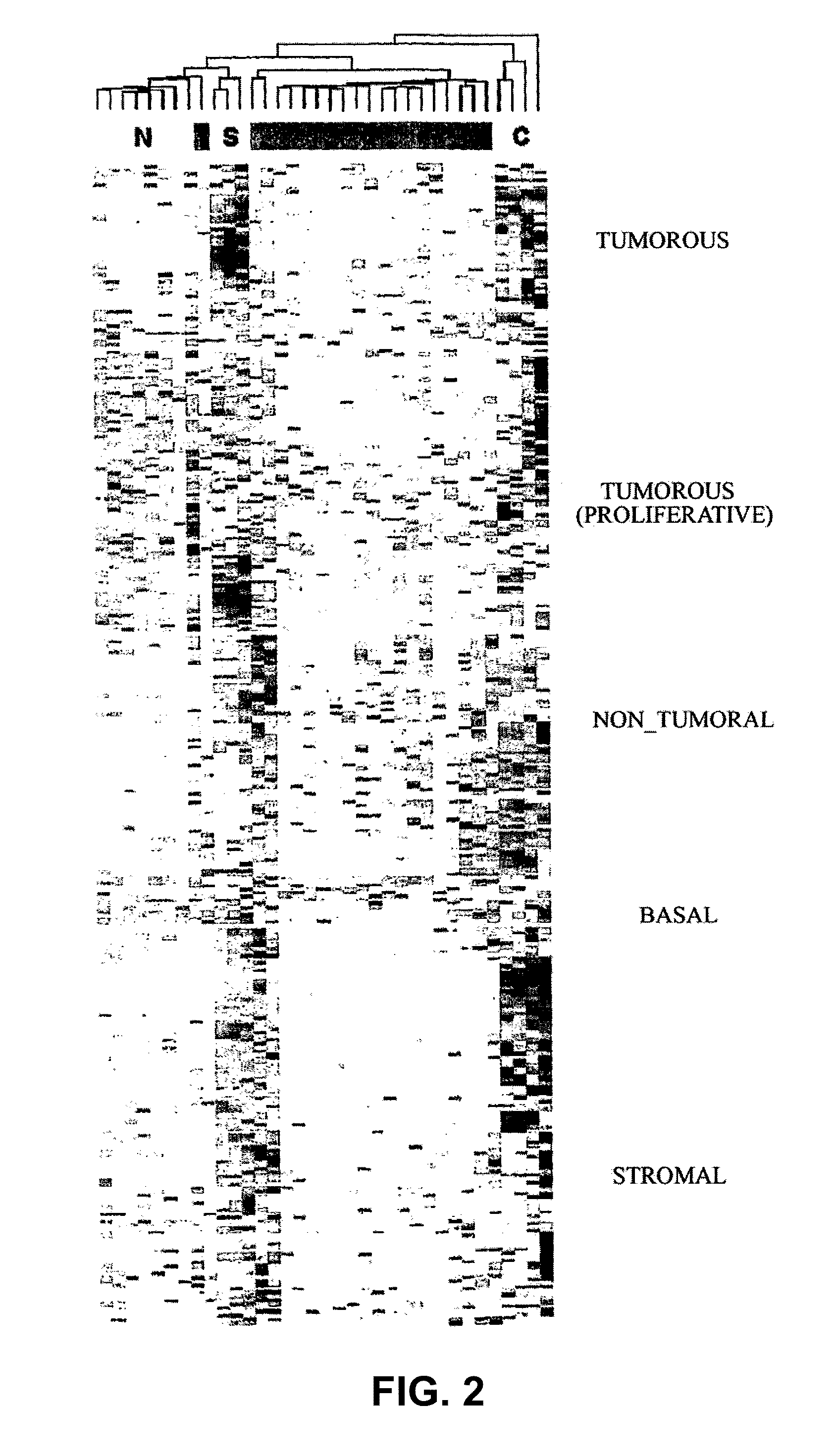
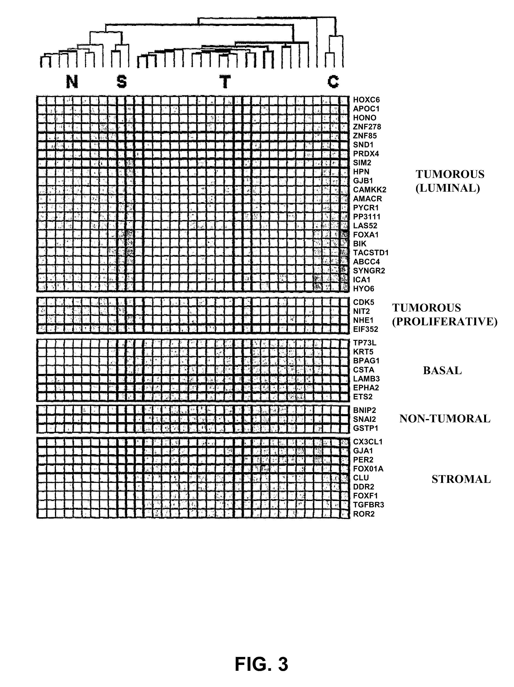
Identification of the Genes Associated with a Cluster Identifying a Prostate Cancer Tumor Pattern

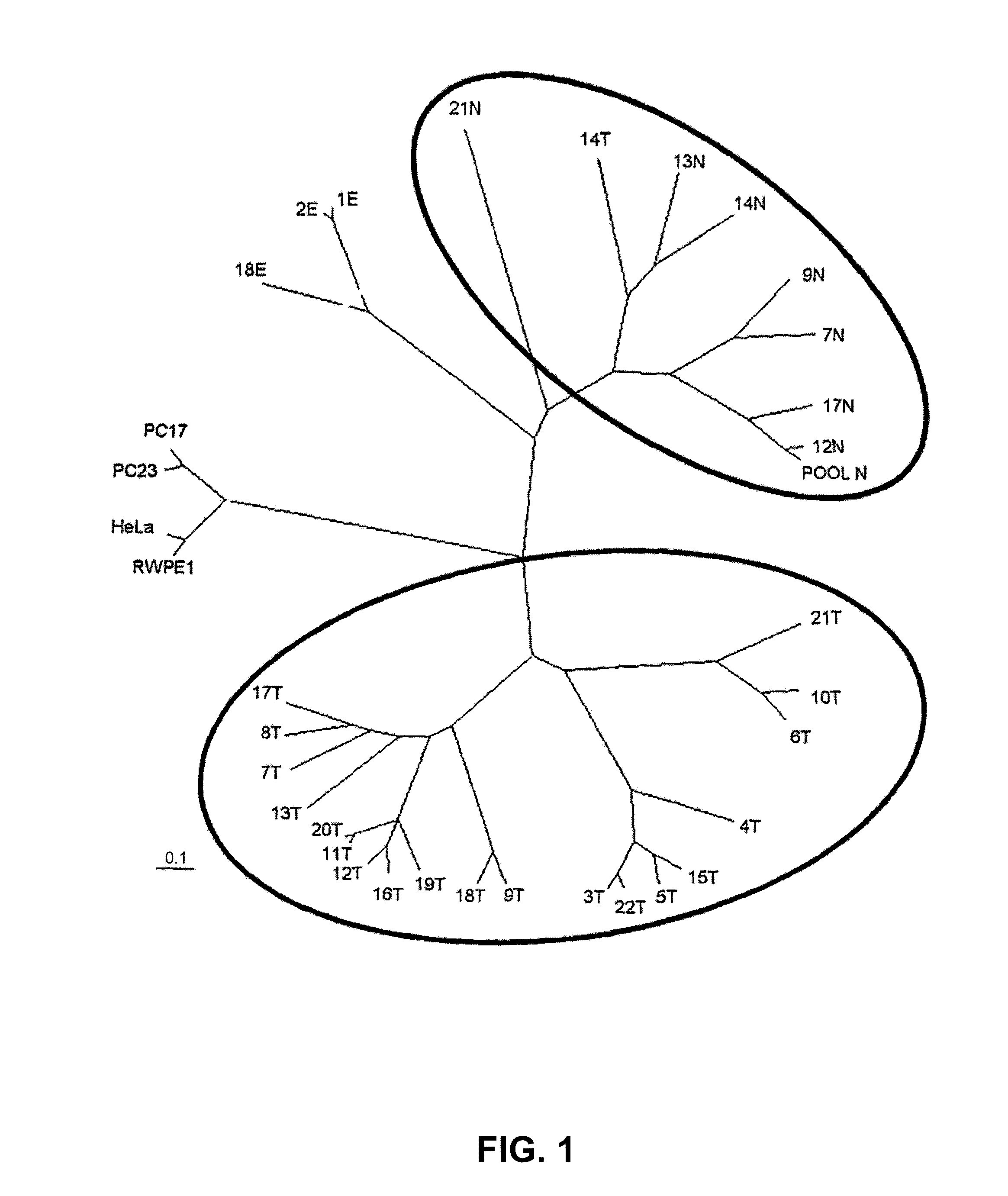
[0074]For the realization of the present invention, a series of 31 human prostate samples were analyzed by hybridization on Affymetrix Human Genome Focus arrays (FIG. 1):

[0075]I. 20 samples enriched with carcinomatous epithelium.

[0076]II. 7 samples enriched with normal epithelium (<1% of cancer cells).

[0077]III. 1 sample comprising a group of 5 normal samples (POOL N).

[0078]IV. 3 samples consisting exclusively of stromal tissue.

[0079]The collected tissues were embedded in OCT, frozen in isopentane, and stored at −80° C. The samples were assessed histologically and selected for analysis in accordance with the following criteria: (a) minimum 90% of pure normal or carcinomatous epithelium in the normal and carcinomatous samples, respectively; (b) absence or minimal presence of foci of inflammation or atrophy. All the samples except three (one normal and two carcinomatous) come from the peri...

example 2

Validation of the Genes Identified as Most Relevant in the Microarray Hybridization Experiments by Means of Real-Time RT-PCR Using the TaqMan LDA Format

[0090]This was done by performing real-time RT-PCR on the genes of greatest interest biologically and as markers from the complete panel of 318 genes identified previously, using both non-microdissected samples and samples laser-microdissected using the PALM instrument. The object of the RT-PCR analysis is to determine the expression levels of these genes in a diagnostic chip-type format, which is smaller and more akin to clinical practice.

[0091]Real-time RT-PCR was carried out for each replica of prostate tissue (in triplicate) or of microdissected samples (in quadruplicate), whether of carcinomatous or normal tissue. Thus, 1 ng of starting total RNA was used for the synthesis of cDNA using the reverse transcriptase Superscript II (Invitrogen) and random hexamers at 42° C. for 50 min, followed by treatment with RNase at 37° C. for 2...

example 3

Immunohistochemical Technique Used on Tissue Microarrays

[0102]The tissue microarrays were constructed using a Beecher instrument (Beecher Instruments) and a 1 mm-diameter needle. Three different microarrays were constructed, containing selected zones of samples of normal prostate, carcinomatous prostate, and PIN tissue, all previously embedded in paraffin. Blocks of lung tissue previously stained with three different colors and placed in different zones of the microarray were used as orientation markers for the samples within the arrays. Complete sections of the microarrays were taken and stained with hematoxylin-eosin to confirm quality. 2 μm thick sections were taken and mounted on xylene-coated glass slides (Dako, Carpinteria, Calif.) for the immunohistochemical stainings. These were done with the Techmate 500 system (Dako), using the Envision system (Dako) for the detection. Briefly, the sections were deparaffinized and rehydrated in graded alcohol series and water. For the dete...


PUM


Abstract

The invention relates to a method for the molecular diagnosis of prostate cancer, comprising the in vitro analysis of the overexpression or underexpression of combinations of genes that can distinguish, with high statistical significance, tumorous prostate samples from non-tumorous prostate samples. The invention also relates to a kit for the molecular diagnosis of prostate cancer, which can perform the above-mentioned detection.

Description

FIELD OF THE INVENTION[0001]The invention falls within the biotechnology sector and specifically within the field of methods for the diagnosis of prostate cancer. Accordingly, the present invention relates to a method for the molecular diagnosis of prostate cancer, comprising the in vitro analysis of the overexpression and underexpression of combinations of genes capable of differentiating between carcinomatous and noncarcinomatous prostate samples with high statistical significance. In particular, the present invention relates to a kit for the molecular diagnosis of prostate cancer capable of carrying out the aforementioned detection.BACKGROUND OF THE INVENTION[0002]Prostate cancer (PC) is a neoplasia having one of the highest rates of mortality and morbidity in industrialized countries and has therefore considerable socioeconomic impact [1], for which reason it is the subject of intensive study. Despite this effort, and in contrast to other types of neoplasia, comparatively little...

Claims


Application Information

Patent Timeline
Patent Type & Authority Applications(United States)
IPC IPC(8): C12Q1/68G01N33/574
CPCG01N33/57434C12Q1/6886C12Q2600/158Y02A90/10C07K16/3069
Inventor THOMSON OKATSU, TIMOTHYGASCON, RAQUEL BERMUDOORTIZ, ANGEL RAMIREZABIA, DAVIDALONSO, CARLOS MARTINEZFERNANDEZ RUIZ, PEDRO LUISFABREGA, BERTA FERRERGUERRI, ELIAS CAMPOVIVES, ELISABET ROSELL
Owner FUNDACIO PRIVADA CLINIC PER A LA RECERCA BIOMEDICA
Why Eureka
Try Eureka