
Human prostate epithelial cell separating and culturing method

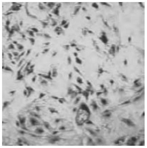
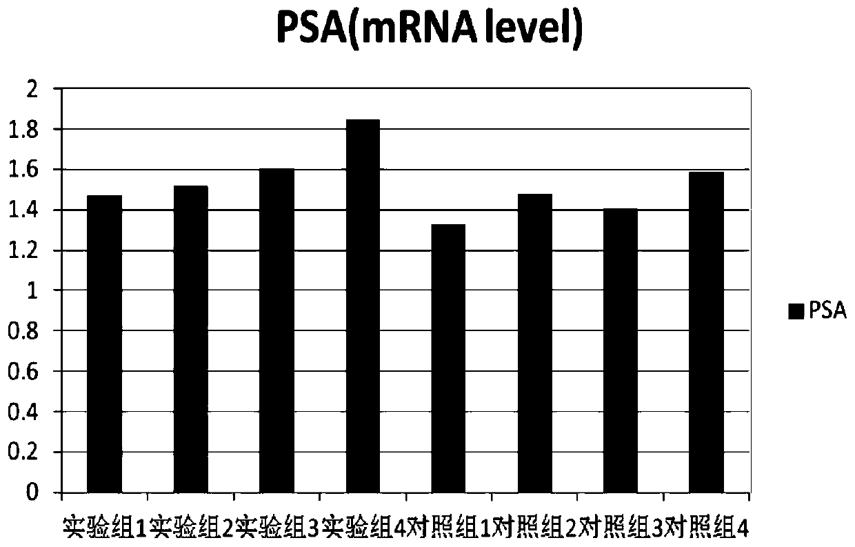
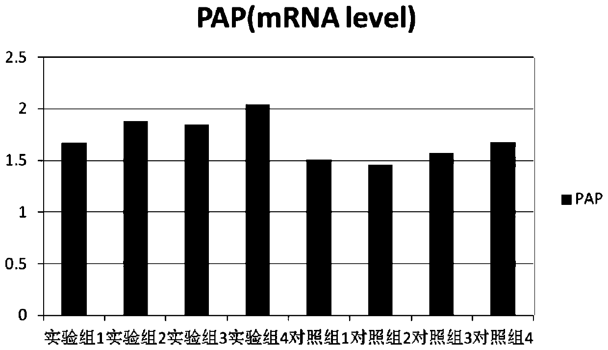
An epithelial cell and culture method technology, applied in the field of separation and culture of human prostate epithelial cells, can solve the problems of immaturity of prostate epithelial cells in vitro, improve cell viability and division ability, increase activity and proliferation rate, and promote cell growth and proliferative effects

Inactive Publication Date: 2019-05-28
北京昱龙摩尔国际生物医学研究院
View PDF1 Cites 0 Cited by
  • Summary
  • Abstract
  • Description
  • Claims
  • Application Information

AI Technical Summary

Problems solved by technology

However, the in vitro culture of prostate epithelial cells is still immature

Method used


Image

  • Human prostate epithelial cell separating and culturing method
  • Human prostate epithelial cell separating and culturing method
  • Human prostate epithelial cell separating and culturing method

Examples


Embodiment 1

[0050] A method for isolating and culturing human prostate epithelial cells, comprising the steps of:

[0051] S1: Sample collection and separation: collect prostate lobe specimens, remove fat tissue, and obtain prostate epithelial tissue;

[0052] S2: Add the digestive solution with double antibody to the prostate epithelial tissue for the first stage of digestion, and cut it into small pieces at the same time; transfer the cut pieces to the culture bottle, add the digestive solution for the second stage of digestion , to obtain dispersed prostate epithelial cells;

[0053] S3: dissolve and disperse the cells with the dispersion liquid, sieve them with 253 μm, 150 μm, 100 μm, and 41 μm nylon meshes sequentially, and collect the residue on the sieve with the dispersion liquid to obtain dispersed prostate epithelial cells;

[0054] S4: Primary culture: adjust the cell concentration of prostate epithelial cells to 4×10 with the primary culture medium 6 cells / ml, cultured in a ca...

Embodiment 2

[0057] A method for isolating and culturing human prostate epithelial cells, comprising the steps provided in Example 1, wherein the specific method of step S2 is as follows:

[0058] S2.1: Add the digestive juice added with double antibody to the prostate epithelial tissue, carry out the first stage of digestion, and cut it into small pieces at the same time, so that it can pass through the No. 14 catheter;

[0059] S2.2: Transfer the shredded pieces to 25 cm with a syringe and 14-gauge catheter 2 In the culture bottle, add digestion solution at a ratio of 1ml per 0.1g of tissue, and place it in a shaking water bath at 37°C for 1 hour to carry out the second stage of digestion;

[0060] S2.3: Centrifuge at 100 g for 5 min to collect dispersed prostate epithelial cells.

[0061] The specific method of step S3 is as follows:

[0062] The cells were redispersed with the dispersion liquid, and sieved with 253, 150, 100, and 41 μm nylon mesh in turn. After each filtration, the s...

Embodiment 3

[0074] A method for isolating and culturing human prostate epithelial cells, comprising the steps provided in Example 2, wherein the digestive solution used is 0.02% EDTA added with 675U / ml collagenase; the double antibody is 100U / ml penicillin and 100 μg / ml ml of kanamycin.


PUM


Abstract

The invention provides a human prostate epithelial cell separating and culturing method. A collected prostate sample is subjected to multiple digestion, dispersion and screening so that other tissuescan be effectively removed to obtain a highly-purified sample; cholera toxin and a hormone are added into a primary culture medium, and thus the cell activity can be effectively improved to promote cell growth so that primary cells of high quality can be obtained; allantoin, creatine and orientin which are added into a passage culture medium can effectively improve the cell vitality and division capability, so that cell growth and proliferation are promoted; 3T3 cells are used as trophoblast, thus a good growth base can be provided for primary prostate epithelial cells, and growth and divisionof the prostate epithelial cells are effectively promoted, so that a culture effect is greatly improved. Through the method, the activity and the proliferation rate of the prostate epithelial cells can be significantly improved, so that high-quality experimental materials are provided for relative research projects.

Description

technical field [0001] The invention belongs to the technical field of tissue engineering, in particular to a method for separating and culturing human prostate epithelial cells. Background technique [0002] The prostate is the largest solid organ among the accessory glands of the male genitalia. Chronic prostatitis, benign prostatic hyperplasia, and prostate cancer are common diseases of the male reproductive system. The pathogenesis is very complicated. Currently, there is no clinically effective treatment. Research on the treatment methods of the above-mentioned diseases needs to deeply explore its pathogenesis. Freshly isolated primary cells retain their original biological genetic characteristics and can reflect the general characteristics of cell growth in vivo, so they are suitable for research on drugs and therapies. The establishment of in vitro cell models of normal prostate epithelial cells and diseased prostate epithelial cells is conducive to a better underst...

Claims


Application Information

Patent Timeline
Patent Type & Authority Applications(China)
IPC IPC(8): C12N5/071
Inventor 张晓南吴芳春吴刘兵陈虎张斌侍晓云
Owner 北京昱龙摩尔国际生物医学研究院
Learn More