Active fixation element

a technology fixing elements, applied in the field can solve the problems of unresolved and somewhat lessened problems relating to the anchoring of active fixation elements in tissue, and achieve the effect of reducing the formation of blood clots

Active Publication Date: 2010-09-16
ST JUDE MEDICAL
View PDF10 Cites 10 Cited by
  • Summary
  • Abstract
  • Description
  • Claims
  • Application Information

AI Technical Summary

Benefits of technology

[0009]However, even by using steroids in connection with implantation the problem associated with anchoring active fixation elements in tissue is not solved, though somewhat lessened. It is therefore a need for an active fixation element that prevents or at least reduces the formation of a blood clot and non-stimulable tissue in the vicinity of the element in the anchored tissue.
[0016]The at least one blood drainage channel will guide and drain blood that is released due to a tissue injury in connection with penetrating and anchoring the fixation element and its connected medical device to the tissue. The blood drainage will reduce the size of any blood and fibrin clot formed from the injury in the vicinity of the fixation element. As the thickness of this non-stimulable clot is reduced, the acute and chronic capture threshold for stimulating the tissue is therefore significantly reduced.
[0021]Allows a faster ingrowth of the active fixation element in the tissue; and
[0022]Can be used in connection with helical wire geometries having improved current density, fixation and stability properties.

Problems solved by technology

However, even by using steroids in connection with implantation the problem associated with anchoring active fixation elements in tissue is not solved, though somewhat lessened.

Method used


Image

  • Active fixation element
  • Active fixation element
  • Active fixation element

Examples


first embodiment

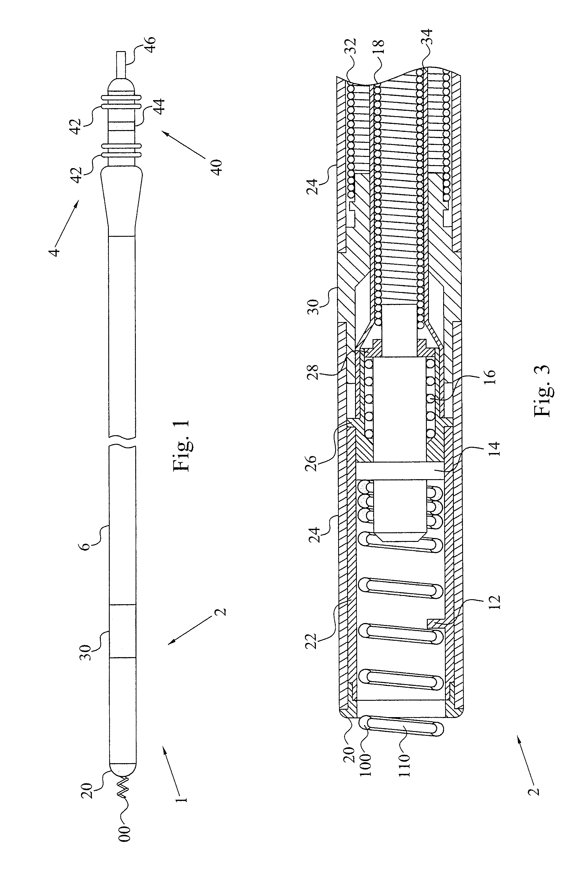
[0065]the helical fixation element is illustrated in FIGS. 4A to 4C. FIG. 4A illustrates a three-dimensional view of the helical fixation element 100. FIG. 4B is a cross-sectional view of the fixation element 100 and FIG. 4C is a cross-sectional view of the helical wire 160 forming the fixation element 100. As is seen in the figures, the fixation element 100 has a general circular cross-section and can therefore be manufactured from a round wire 160. However, in clear contrast to the prior art fixation elements, a blood drainage channel 110 in the form of a groove or recesses 110 in the helical wire 160 runs along the helical windings 120 from the distal helix end 140 to the proximal lead end 150.

[0066]In the figures, the groove 110 is running along the windings 120 and faces radially out from an inner diameter space 130 confined by the helix windings 120. The present invention is though, no limited to such a relative channel positioning in the wire 160. In an alternative embodiment...

second embodiment

[0070]Compared to the helix embodiment disclosed by FIGS. 4A to 4C, the second embodiment has a better penetration design that is more suited for penetrating the endocardium and the myocardium while reducing the caused tissue injury. The helical fixation element also has improved ability to withstand pull forces on the distal lead end when the helix is fully extended in the tissue. Since the crescent-shape implies the formation of edges on the windings 220, the current density increases at the edges, thereby allowing a reduction in the energy consumption in connection with tissue stimulation.

third embodiment

[0071]a helical fixation element 300 according to the present invention is illustrated in FIGS. 6A to 6C. Compared to the previous embodiments, the cross-sectional dimension of the helix wire 360 is smaller in the direction of the longitudinal axis of the fixation element 300 than in a direction transverse to the longitudinal axis. The helical wire 360 can have a general rectangular cross-section with a drainage channel running along the helix windings 320 from the tissue-penetrating end 340 and preferably up to the device-connecting end 350. The channel 310 is preferably formed in at least one of the longer sides of the rectangular, implying that the channel 310 is facing the device-connecting 350 or the tissue-penetrating 340 end of the fixation element 300.

[0072]Instead of utilizing a rectangular cross-section, the wire 360 can be elliptical with the major axis perpendicular to the longitudinal helix axis. FIGS. 6A to 6C illustrate such an embodiment with the drainage channel 310...


PUM


Abstract

A helical fixation element of an implantable medical lead. The fixation element has at least one blood drainage channel running along at least a tissue-penetrating portion of the helix windings of the fixation element. The channel guides, during penetration and anchoring of the fixation element and the lead in a tissue, blood leaking from the tissue away from the vicinity of the fixation element, thereby reducing the size of a fibrin clot formed around the fixation element. The capture threshold for stimulating the tissue is therefore reduced.

Description

BACKGROUND OF THE INVENTION[0001]1. Field of the Invention[0002]The present invention generally relates to active fixation elements, and in particular such fixation elements for usage in implantable medical leads.[0003]2. Description of the Prior Art[0004]Body implantable electrical leads form the electrical connection between an implantable medical device (IMD), such as cardiac pacemaker or cardiac defibrillator, and body tissue, such as the heart, which is to be electrically stimulated. As is well known, the leads connecting the IMD with the tissue may be used for pacing / defibrillation and for sensing electrical signals produced by the tissue.[0005]The implantable leads of today and in particular cardiogenic implantable leads can generally be divided into two classes depending on the tissue anchoring arrangement of the leads. Firstly, so-called passive fixation leads comprise radially protruding elements in the distal lead ends. These elements can become embedded in the trabecular...

Claims


Application Information

Patent Timeline
Patent Type & Authority Applications(United States)
IPC IPC(8): A61N1/18
CPCA61N1/0573
Inventor JARL, PER E.HILL, ROLFSTEGFELDT, OLOF
Owner ST JUDE MEDICAL
Why Eureka
Try Eureka