
Biological valve for venous valve insufficiency

a technology of venous valve and biological valve, applied in the field of biological valve, can solve the problems of limited venous valve replacement and therapy, eliciting a deleterious immune response,

Inactive Publication Date: 2010-09-16
HANCOCK JAFFE LAB INC
View PDF54 Cites 11 Cited by
  • Summary
  • Abstract
  • Description
  • Claims
  • Application Information

AI Technical Summary

Benefits of technology

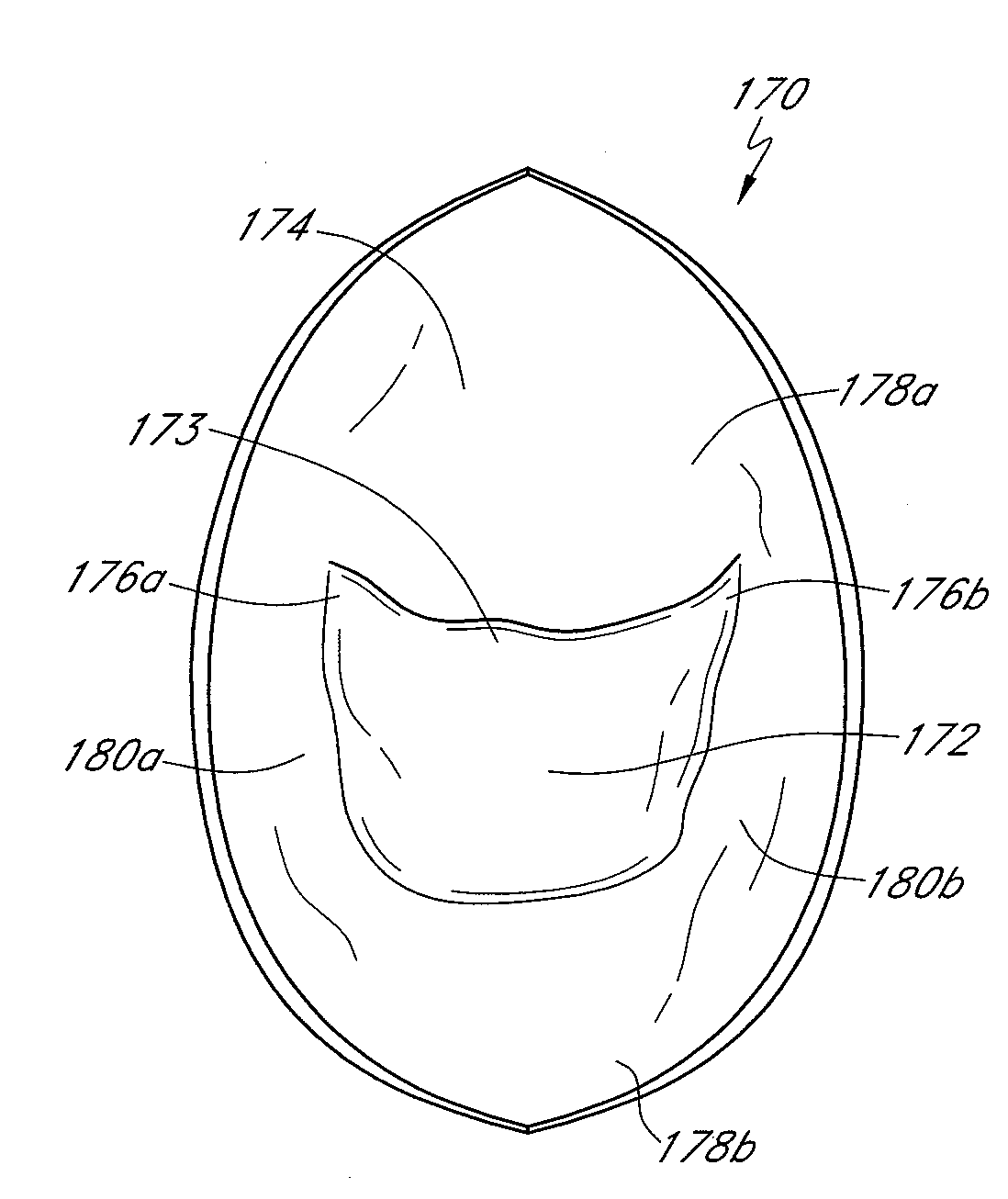
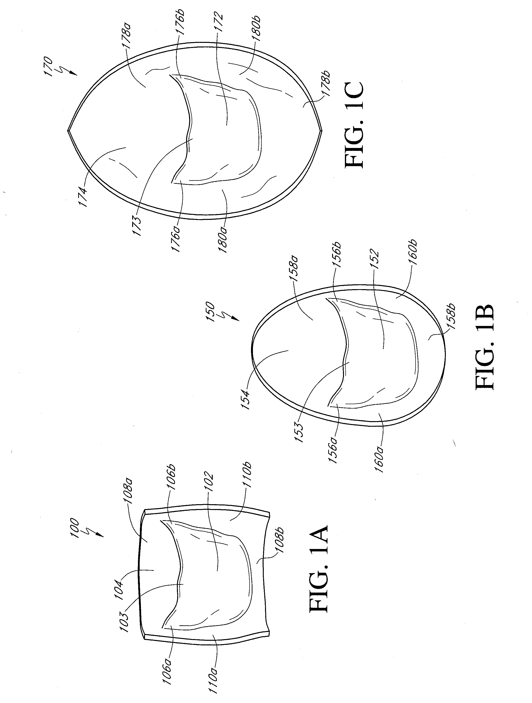
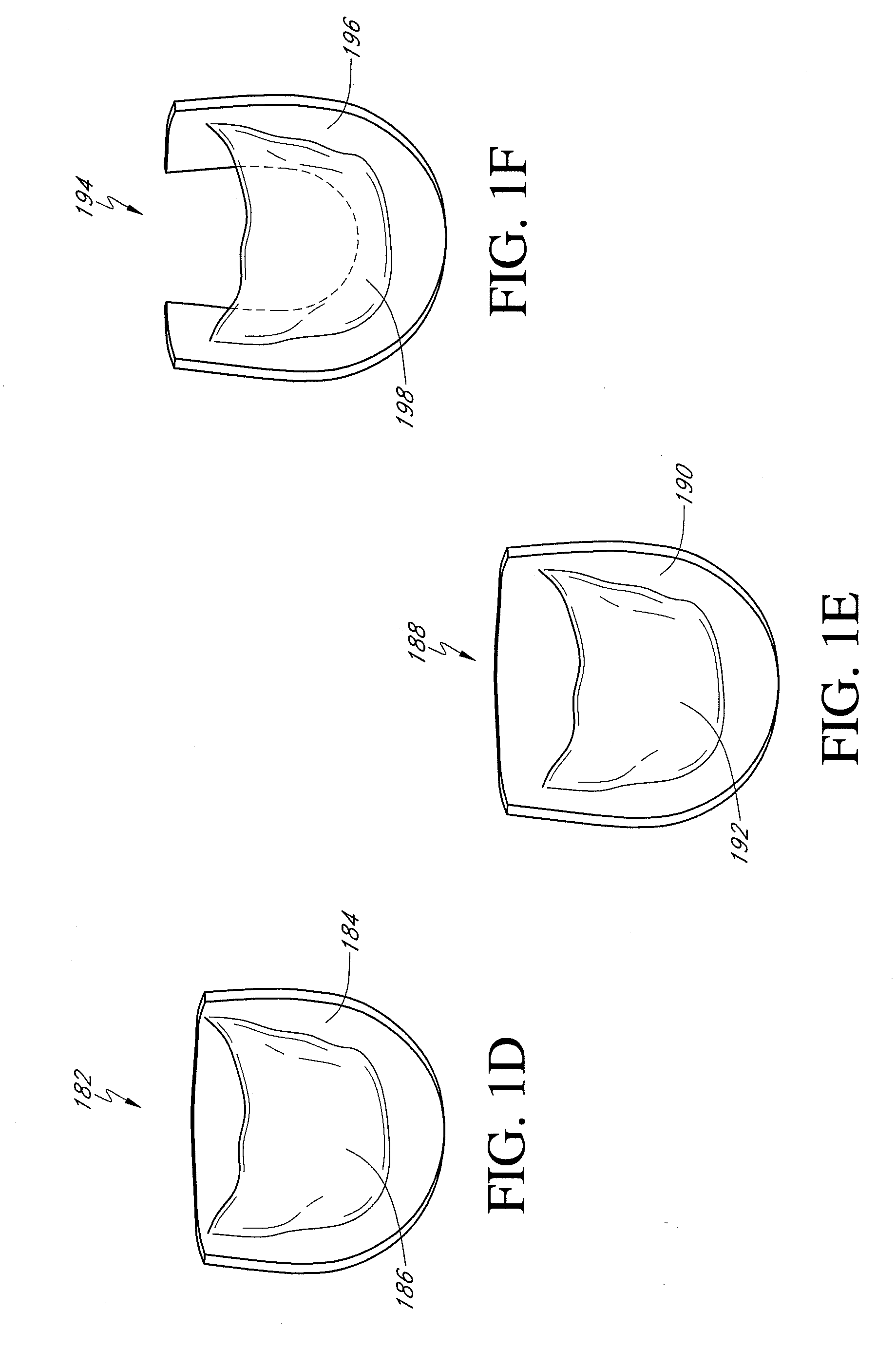
"The patent describes a bioprosthetic valve made from a xenogeneic source (often a porcine heart valve) that can be implanted into a host vein of a subject. The valve consists of a single leaflet attached to a patch of valve wall tissue. The patch can be a portion of the heart valve or a separate segment of valve wall tissue. The patch can extend circumferentially on either side of the leaflet or axially above and below the leaflet. The valve can be fixed using a fixation treatment, such as exposure to glutaraldehyde solution. The patent also describes a method of manufacturing the replacement valve and a method of treating a malfunctioning valve in a host vein. The technical effects of the patent include providing a bioprosthetic valve that can be implanted into a host vein of a subject, providing a method of manufacturing the valve, and providing a method of treating a malfunctioning valve."

Problems solved by technology

Currently, venous valve replacement and therapy is limited to allograft or cryopreserved valves.
Allograft valves are difficult to harvest and cryopreserved valves can elicit a deleterious immune response.

Method used


Image

  • Biological valve for venous valve insufficiency
  • Biological valve for venous valve insufficiency
  • Biological valve for venous valve insufficiency

Examples


example

Hemodynamic Evaluation of Valve Device

[0053]To evaluate the hydrodynamic performance and leaflet motion characteristics of a venous valve device according to the disclosure, several valve devices of various sizes were constructed and tested in the aortic chamber of a pulsatile flow heart valve test apparatus. Hydrodynamic performance was observed under a range of conditions typical of the upper leg of a human being. Leaflet function (i.e., opening and closing) was confirmed for all valve devices under all test conditions studied.

Construction of Valve Devices

[0054]Three unconstrained diameters (10 mm, 12 mm, and 14 mm) believed to be suitable for valve devices intended to be implanted in a human vein were selected for evaluation. For each unconstrained diameter, three valve devices were constructed by attaching a gluteraldehyde crosslinked bioprosthetic valve to a support frame by suturing. All specimens were submerged in saline following construction and subjected to irradiation.

Sim...


PUM


Abstract

A bioprosthetic valve for repairing a deep venous insufficiency in a subject includes a single leaflet from a xenogeneic heart valve attached at natural margins of attachment to a patch of valve wall tissue. The patch may extend axially above and below the leaflet and circumferentially on either side of the leaflet to provide a region for attaching the patch to a fenestration in a host vein. A bioprosthetic valve may be manufactured by excising a portion of a xenogeneic heart valve including a single leaflet and contiguous wall tissue, and may further comprise shaving off excess leaflet tissue from adjacent leaflets. A method of replacing a malfunctioning venous valve in a subject includes providing a bioprosthetic valve as described above and inserting it to the host vein.

Description

CROSS-REFERENCE TO RELATED APPLICATIONS[0001]This application is a divisional of U.S. application Ser. No. 12 / 252,253, filed Oct. 15, 2008, which claims the benefit of U.S. Provisional Application No. 60 / 980,708, filed Oct. 17, 2007, the entire disclosure of each of which is hereby incorporated by reference.BACKGROUND OF THE INVENTION[0002]1. Field of the Invention[0003]This application generally relates to a biological valve for the replacement of a venous valve of the deep venous system of the leg or other veins of comparable caliber.[0004]2. Description of the Related Art[0005]Patients with chronic deep vein insufficiency may require the repair or replacement of at least one of the venous valves of the deep veins of the leg. Also, in some cases the venous valve replacement may involve the iliac veins.[0006]Present therapy involves either the surgical repair of the valve leaflets or external banding of the vein to decrease the diameter and bring the weakened leaflets to a geometry...

Claims


Application Information

Patent Timeline
Patent Type & Authority Applications(United States)
IPC IPC(8): A61F2/06
CPCA61F2/2412A61L27/507A61L27/3625A61F2/2475A61F2/2415
Inventor JAFFE, NORMAN
Owner HANCOCK JAFFE LAB INC
Learn More