Patient registration system

a registration system and patient technology, applied in the field of patient registration system, can solve the problems of invasiveness to the body, displacement of body markers, and inability to embed markers in the body of patients, and achieve the effect of high accuracy and short tim

Inactive Publication Date: 2010-09-30
MITSUBISHI ELECTRIC CORP
View PDF1 Cites 28 Cited by
  • Summary
  • Abstract
  • Description
  • Claims
  • Application Information

AI Technical Summary

Benefits of technology

[0016]It is an object of the present invention is to provide a patient registration system which can adequately estimate an amount of displacement not only on an imaging plane but also outside the imaging plane by analyzing in advance both of CT data and a DRR image during DRR image generation process, thereby achieving registration of patient in a short time and with high accuracy.
[0057]According to an embodiment of the present invention, by carrying out the processes of extraction of the three-dimensional characteristic, characteristic evaluation and area limitation, an amount of displacement not only on an imaging plane but also outside the imaging plane can be adequately estimated during patient registration. Further, the amount of displacement can be estimated within the area of interest, thereby achieving registration of patient in a short time and with high accuracy.

Problems solved by technology

However, in a case in which a long period of time has elapsed from the CT scanning to the radiotherapy, a position and a body posture of the patient lying on a treatment table during treatment are often different from these of the patient during planning of treatment.
According to the above existing technique, however, the markers acting as indicators for registration may form blind spots in the X-ray television image captured using an X-ray television imaging device.
However, embedding body markers in a body of patient will cause not only a problem of invasiveness to the body but also another problem of displacement of the body markers after a long period of time has elapsed from CT scanning to radiotherapy.
Moreover, in some cases, it is not possible to embed the markers in the body of patient depending on conditions of the diseased site.
This approach can calculate a rotation on a two-dimensional imaging plane, but it is difficult to calculate a rotation outside the imaging plane as an amount of three-dimensional correction.

Method used


Image

  • Patient registration system
  • Patient registration system
  • Patient registration system

Examples


embodiment 1

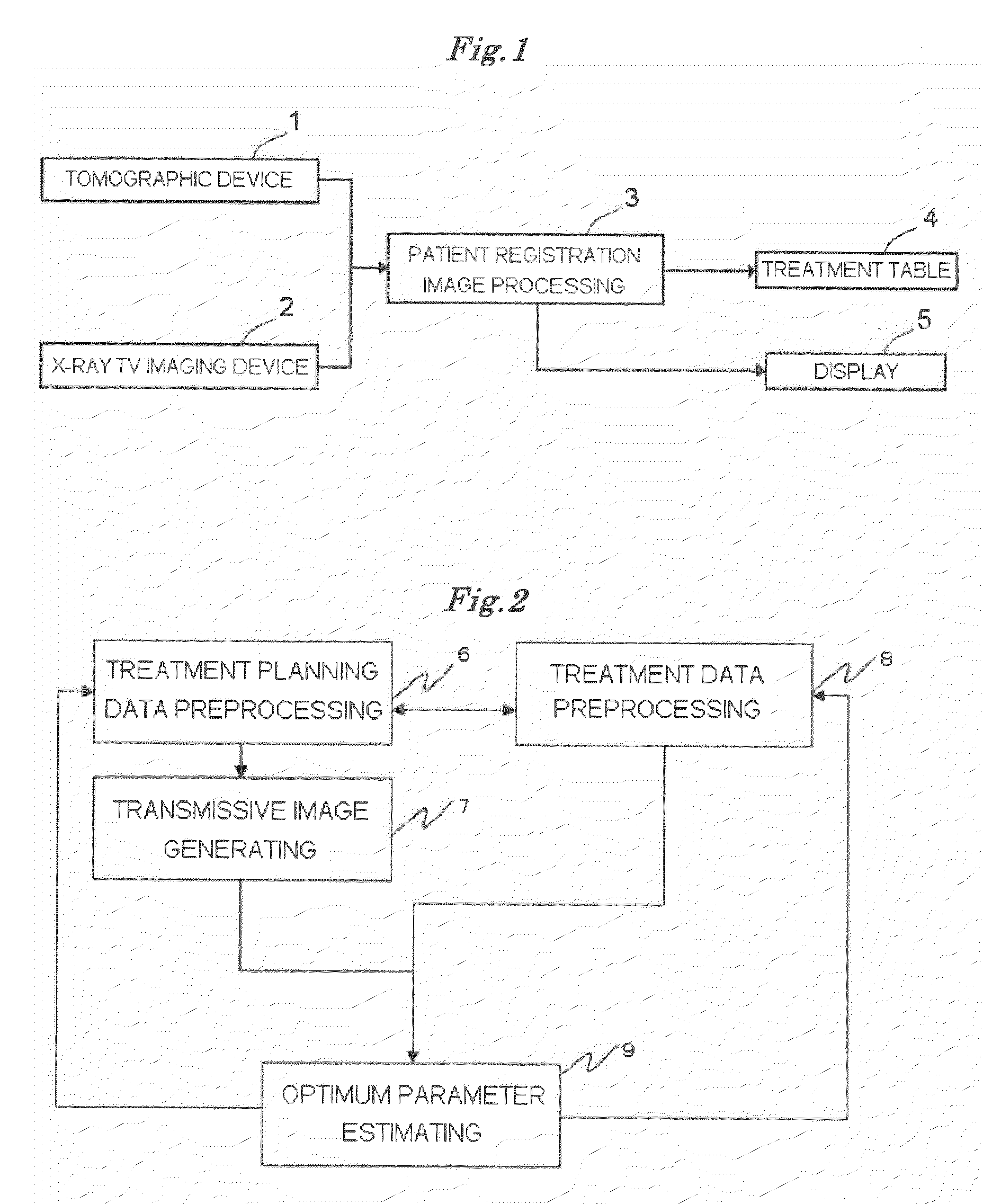
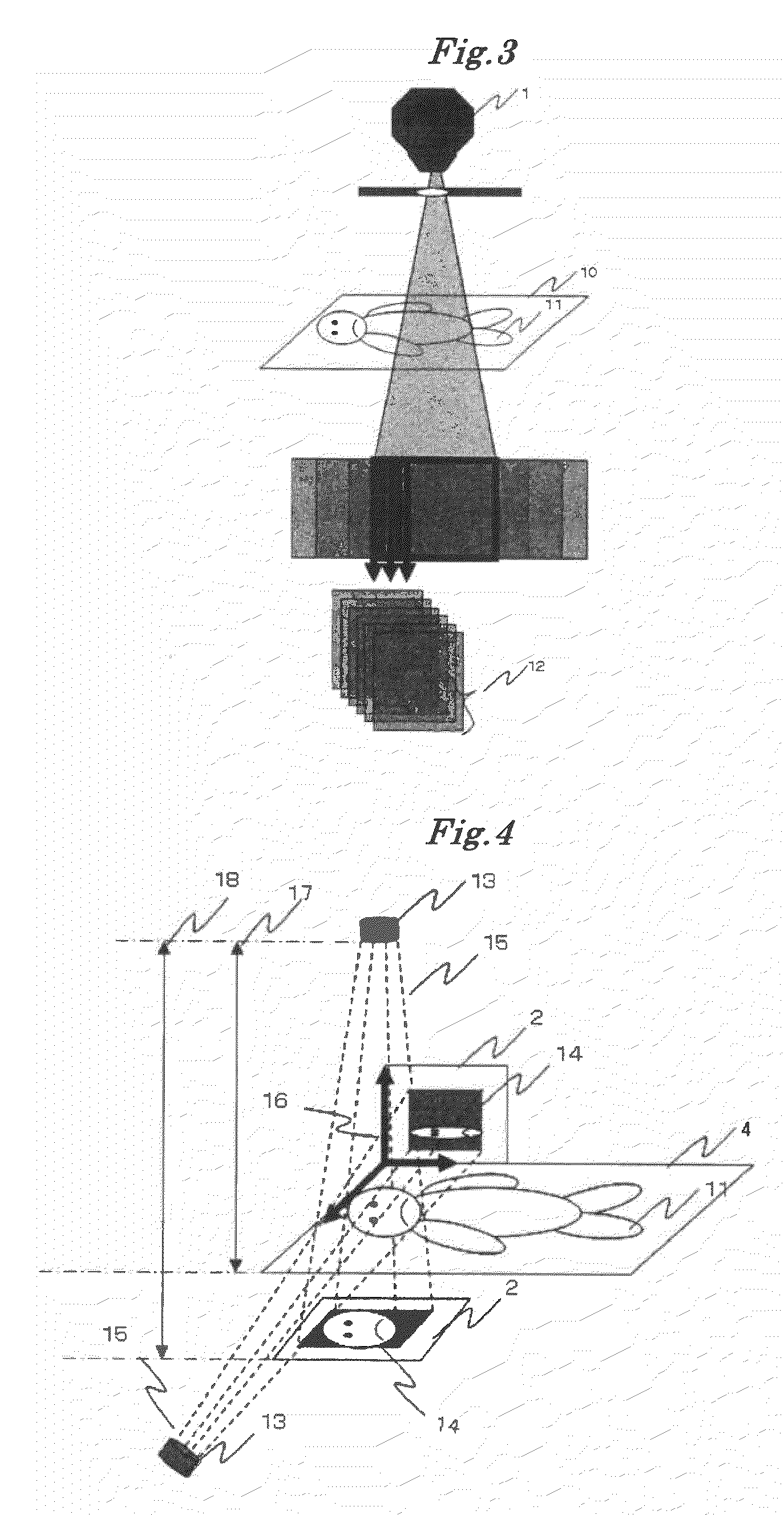
[0074]FIG. 1 shows a configurational view illustrating a patient registration system according to a first embodiment of the present invention. The patient registration system is configured of a tomographic device 1, an X-ray television imaging device 2, a patient registration image processing device 3, a treatment table 4, a display device 5, and an input device (not shown) such as keyboard, mouse and the like.

[0075]The tomographic device 1 is configured of, for example, an X-ray CT (Computed Tomography) scanner, and serves a function of capturing 3-D (three-dimensional) CT data of a diseased site. The X-ray television imaging device 2 is configured of, for example, an X-ray image intensifier tube, and serves a function of capturing an X-ray television image of the diseased site. The X-ray television imaging device 2 is usually installed integrally with a radiotherapy equipment.

[0076]The patient registration image processing device 3 can be configured of one or more computers and th...

embodiment 2

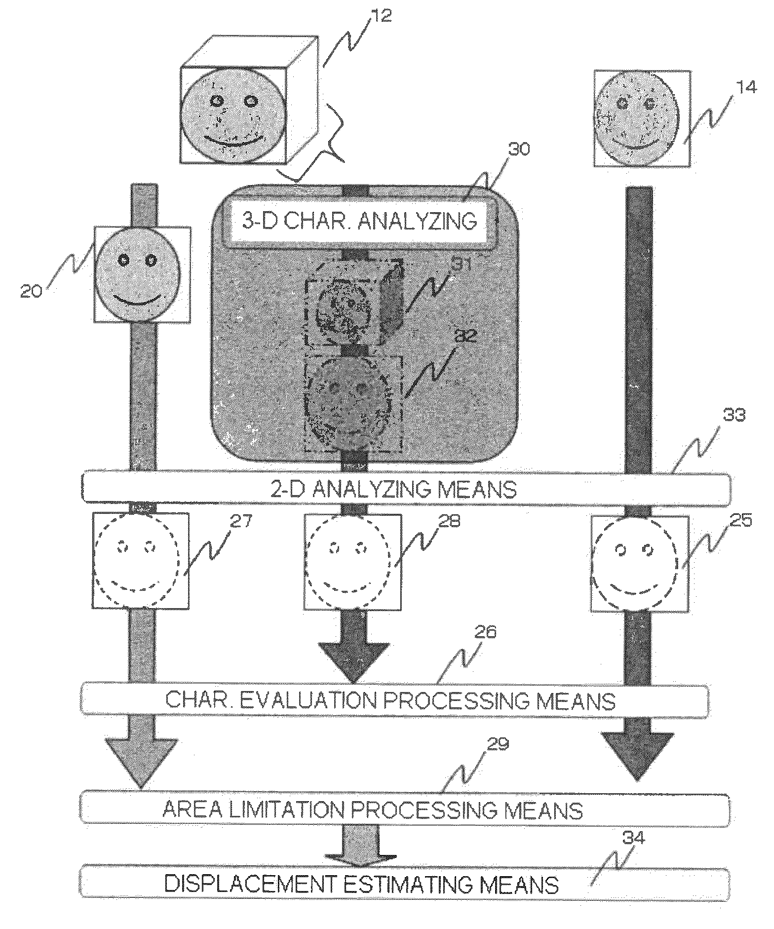
[0099]In this embodiment, in the process flow shown in FIG. 6 of Embodiment 1, the data is compressed to calculate the amount of displacement. This processing compresses the data without damaging the three-dimensional characteristic of the CT data 12. Then, registration is carried out using the compressed data. For this method of compression without damaging the three-dimensional characteristic, the three-dimensional characteristic analyzing means 30 is used.

[0100]This method is carried out, as described in Patent Publication 4, by expanding the processing of the two-dimensional image as shown in FIG. 7 to the three-dimensional volume data as shown in FIG. 8. First, the whole CT data 12 is divided into blocks, and then compressed using high resolution volume data 39 and low resolution volume data 40 that are areas before and after compressing the divided area, without damaging the three-dimensional characteristic. Alternatively, the CT data 12 may be compressed without carrying out ...

embodiment 3

[0105]In this embodiment, in the process flow shown in FIG. 6 of Embodiment 1, the displacement estimation is carried out using only the characteristic amount obtained by the characteristic evaluation processing means 26, without using the three-dimensional characteristic analyzing means 30, the characteristic evaluation processing means 26, and the area limitation processing means 29. After convergence, the amount of displacement is varied as the parameter for out-of-plane rotation.

[0106]The parameter is varied by the following method. In a generation method here, the parameters are varied at random (Mersenne twister or the like). Alternatively, the parameters are varied by changing a varying range of parameter at an arbitrary step such as (+5 degrees to −5 degrees) with respect to an out-of-plane rotation axis. In this case, limiting values of the varying range is maximum values of movement of the treatment table corresponding to the out-of-plane rotation. Alternatively, the param...


PUM


Abstract

There is provided a patient registration system according to the present invention, including: a CT data capturing device; an X-ray television imaging device; and an image processing device for generating a two-dimensional DRR image based on the captured CT data, and then calculating an amount of displacement between a first diseased site position when the CT data is captured and a second diseased site position when the X-ray television image is captured. The image processing device carries out processes of: three-dimensional analysis for extracting an amount of three-dimensional characteristic from the three-dimensional CT data; two-dimensional analysis for extracting an amount of two-dimensional characteristic from both of the DRR image and the X-ray television image; characteristic evaluation for evaluating the extracted characteristic amounts; area limitation for selecting an area where the evaluated characteristic amounts are present; and displacement estimation for estimating an amount of displacement between the first diseased site position and the second diseased site position within the selected area, whereby achieving quick and accurate registration of patient.

Description

BACKGROUND OF THE INVENTION[0001]1. Field of the Invention[0002]The present invention relates to a patient registration system which is suitably used for radiotherapy in which a nidus of a patient can be treated by irradiating the nidus with a radiation beam or a particle beam.[0003]2. Description of the Related Art[0004]A typical patient registration system initially receives 3-D (three-dimensional) CT (Computed Tomography) data for planning of treatment, which can be captured by scanning a nidus of a patient using a tomographic equipment, such as X-ray CT scanner. Planning of treatment can be developed based on diagnostic results of the CT data. At this time, positions and shapes of tumor sites are identified based on the 3-D CT data to determine irradiating directions and doses of radiation.[0005]Next, radiotherapy will be carried out according to the determined plan of treatment. However, in a case in which a long period of time has elapsed from the CT scanning to the radiothera...

Claims


Application Information

Patent Timeline
Patent Type & Authority Applications(United States)
IPC IPC(8): G06K9/62H05G1/60
CPCA61B6/032A61N2005/1062A61N5/1049A61B6/5235
Inventor YAMAKOSHI, RYOICHIHIRASAWA, KOSUKEOKUDA, HARUHISAKAGE, HIROSHISUMI, KAZUHIKOSAKAMOTO, HIDENOBU
Owner MITSUBISHI ELECTRIC CORP
Why Eureka
Try Eureka