System for Multi Nucleus Cardiac MR Imaging and Spectroscopy

a multi-nucleus, mr imaging technology, applied in the direction of nmr measurement, instruments, applications, etc., can solve the problems of reduced spectral resolution and contamination of the 1h mr spectrum, affecting the reliability of myocardial 1h mr spectroscopy, and relatively long total examination tim

Active Publication Date: 2010-10-21
SIEMENS MEDICAL SOLUTIONS USA INC
View PDF4 Cites 30 Cited by
  • Summary
  • Abstract
  • Description
  • Claims
  • Application Information

AI Technical Summary

Problems solved by technology

Performing clinical cardiac spectroscopy involves a number of problems.
Total examination time is relatively long, and motion artifacts including signal contamination by surrounding tissue (chest wall, blood pool) require correction.
Motion artifacts from cardiac and respiratory motion have a negative effect on the reliability of myocardial 1H MR spectroscopy.
Motion of the heart relative to the volume of interest may lead to reduced spectral resolution and contamination of the 1H MR spectrum by, for example, epicardial fat.
In addition, respiratory motion may negatively influence 1H MR spectral resolution by preventing optimal shimming and water suppression.

Method used


Image

  • System for Multi Nucleus Cardiac MR Imaging and Spectroscopy
  • System for Multi Nucleus Cardiac MR Imaging and Spectroscopy
  • System for Multi Nucleus Cardiac MR Imaging and Spectroscopy

Examples


Embodiment Construction

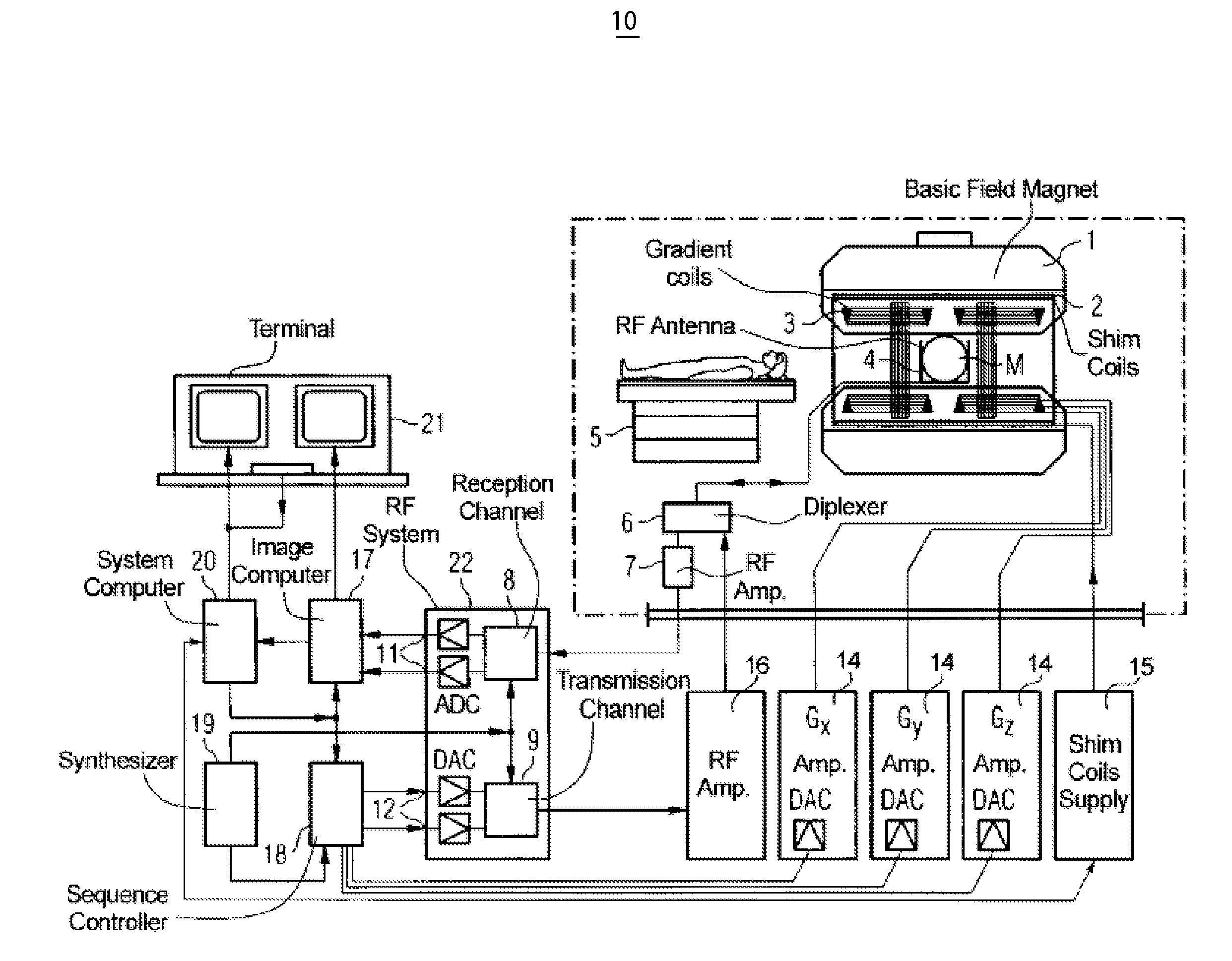
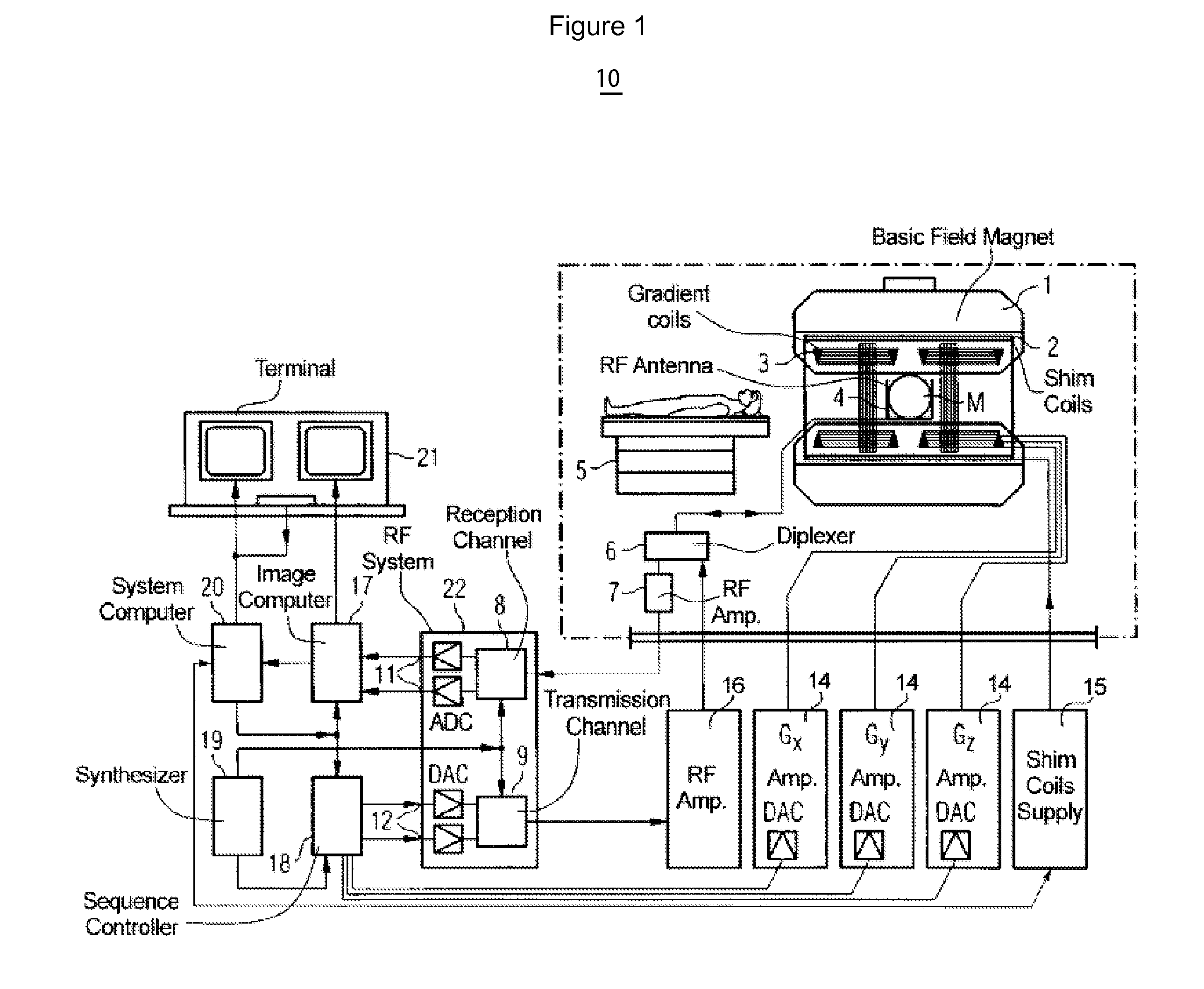
[0013]Respiratory motion compensation using one dimensional / two dimensional (1D / 2D) PACE (Prospective Acquisition Correction) based on echoes from designated lines (1D) or areas (2D) termed navigators is known to be used for double-triggered cardiac proton spectroscopy. The double triggering comprises triggering on both a respiratory cycle PACE derived signal and on a cardiac e.g., ECG signal. The navigators measure the displacement of the liver-lung interface during free breathing. The displacement information allows for double triggering on a defined window within the respiratory cycle and on a defined trigger delay after an R-wave based on an ECG signal. Furthermore, the displacement information allows the excitation volume to be shifted by the determined respiratory displacement within the defined window in real-time (volume tracking). Therefore, application of respiratory navigator gating and tracking improves spectral resolution and reproducibility for metabolic imaging of myo...


PUM


Abstract

A system for respiratory motion compensated MR imaging or spectroscopy, comprises an MR imaging system. The MR imaging system performs a single imaging scan including, acquiring a first imaging data set representing a spatially localized first imaging region located on a patient diaphragm, using a first RF excitation pulse sequence and by transmitting a nuclei excitation first resonant frequency and receiving data substantially at the first resonant frequency. The MR imaging system derives data representing diaphragm position over a respiratory cycle using the first imaging data set, in the single imaging scan. The MR imaging system in response to determining the diaphragm position is within a predetermined window, acquires a second anatomical imaging data set representing a spatially localized second imaging region using a second RF excitation pulse sequence and by transmitting a nuclei excitation second resonant frequency different to the first resonant frequency and receiving data substantially at the second resonant frequency in the single imaging scan.

Description

[0001]This is a non-provisional application of provisional application Ser. No. 61 / 169,536 filed 15 Apr. 2009, by Jian Xu.FIELD OF THE INVENTION[0002]This invention concerns an MR imaging system for respiratory motion compensated MR imaging or spectroscopy, using first and second different RF frequencies to acquire in a single scan, imaging data sets representing a first region used for tracking respiratory movement and a second region for spectroscopy, for example.BACKGROUND OF THE INVENTION[0003]MR spectroscopy (MRS) is used for the non-invasive study of cardiac metabolism without the need for the application of external radioactive tracers, such as involved in Positron Emission Tomography (PET). Nuclei of interest for metabolic MRS studies include 1H, 13C, 19F, 23Na, 31P, 39K, 87Rb. Both 31P and 1H-MRS-studies are of clinical interest. Specifically, 1H-MRS is used to measure total creatine and to evaluate the oxygenation of cardiac tissue or to evaluate diet and therapy effects. ...

Claims


Application Information

Patent Timeline
IPC IPC(8): G01R33/48
CPCA61B5/055A61B5/0816A61B5/7285G01R33/5676G01R33/5607G01R33/563G01R33/5673G01R33/5602
Inventor XU, JIAN
Owner SIEMENS MEDICAL SOLUTIONS USA INC
Why Eureka
Try Eureka