Computer-aided detection (CAD) of a disease

a disease and computer technology, applied in the field of computer-aided detection of diseases, to achieve the effect of reducing the number of patients

Inactive Publication Date: 2010-10-21
KONINKLIJKE PHILIPS ELECTRONICS NV
View PDF11 Cites 10 Cited by
  • Summary
  • Abstract
  • Description
  • Claims
  • Application Information

AI Technical Summary

Benefits of technology

[0008]Accordingly, the invention preferably seeks to mitigate, alleviate or eliminate one or more of the above mentioned disadvantages singly or in any combination. In particular, it may be seen as an object of the present invention to provide a method for easier and / or faster analysis of a medical image data set for likelihood for a disease.
[0013]The invention is particularly, but not exclusively, advantageous for obtaining an improved computer-aided detection (CAD) method facilitating that more efficient computations can be performed because the degree of analysis in a certain region of the part of the patient, e.g. the lung, can be adjusted or tailored to the level of probability of the disease in the region. Thus by application of the present invention it is possible to increase computational speed and thereby diseases like cancer, in particular cancer nodules in the lungs, can be more effectively found from medical image analysis.
[0016]Optionally, the step a) of segmenting the medical image data set using an anatomical model may comprise as an input a position dependent probability (P_r) for the disease. Thus, segmentation resolution may be fine or coarse depending on the level of position dependent probability (P_r). In particular, one or more regions of the medical image data set may not be segmented due to the position dependent probability (P_d) for the disease in the one or more regions being below a predetermined threshold value thereby excluding regions with quite low probability in order to simplify further processing and computations.

Problems solved by technology

However, this is a preliminary result or final result of the method and is not used as an input to simplify subsequent calculations during the CAD process.

Method used


Image

  • Computer-aided detection (CAD) of a disease
  • Computer-aided detection (CAD) of a disease
  • Computer-aided detection (CAD) of a disease

Examples


Embodiment Construction

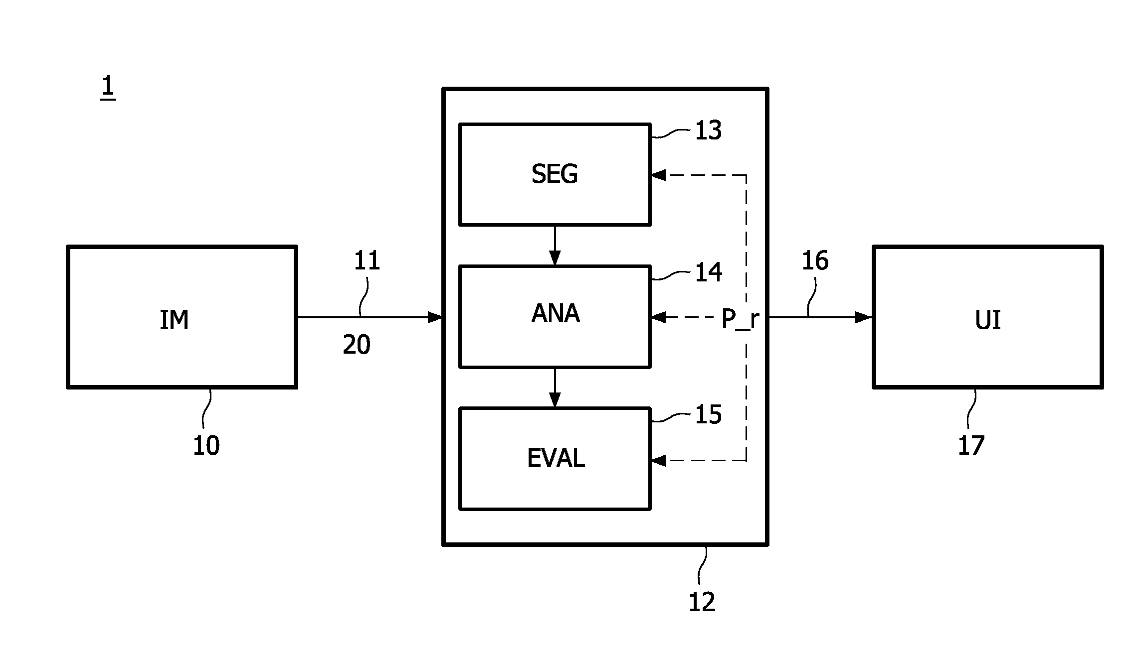
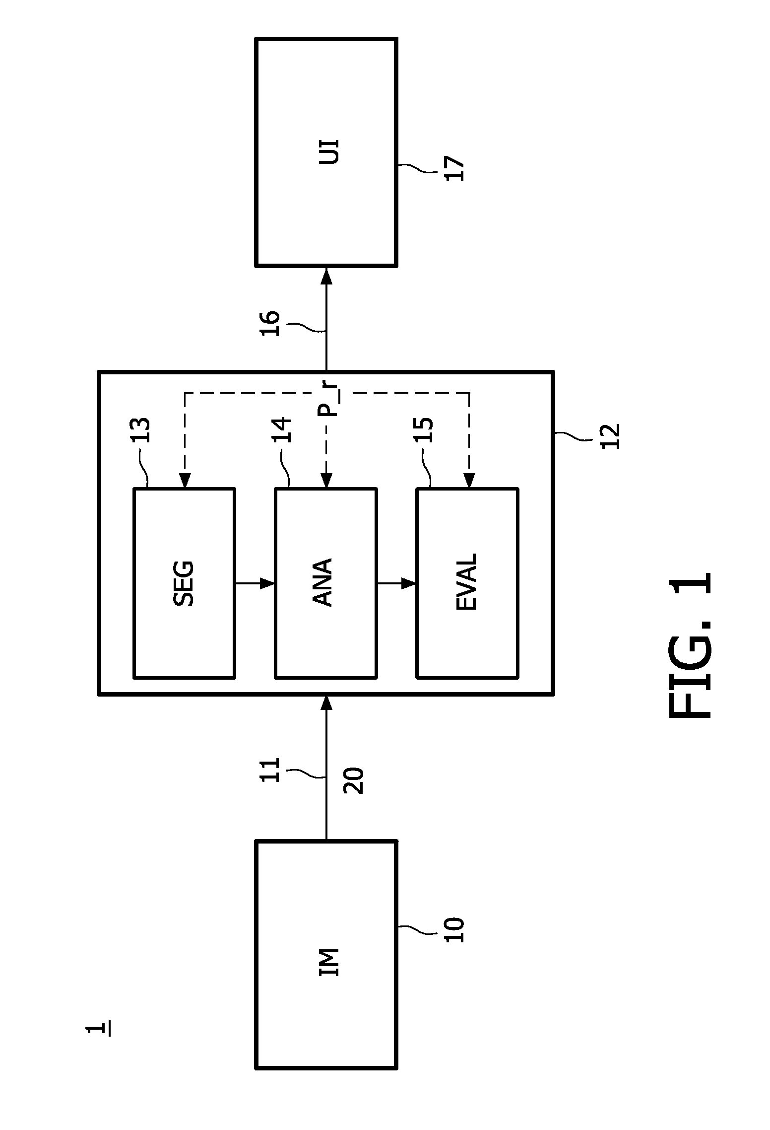
[0039]FIG. 1 schematic drawing of a combined imaging modality and computer system 1 for one embodiment of the present invention. The computer system 1 being arranged for performing CAD from a medical image data set 20 obtained from a medical imaging modality IM, such as computer tomography (CT), magnetic resonance imaging (MRI), positron electron tomography (PET), single photon emission computed tomography (SPECT), ultrasound scanning, and rotational angiography, or any other medical imaging modalities. The transmission from the modality IM to the unit 12 can be via a dedicated connection means 11 (short range or long range, possibly via internet) or by wireless transmission.

[0040]The unit 12 of the computer system 1 is arranged for performing computer-aided detection (CAD) of a disease on a medical image data set 20. Segmentation means 13 are provided for segmenting the medical image data set 20 using an anatomical model, preferably an augmented model. For general reference to segm...


PUM


Abstract

The present invention relates to a method for performing computer-aided detection (CAD) of a disease, e.g. lung tumours, on a medical image data set (20) from a imaging modality, such as MRI or CT. Initially, there is perform a segmentation of the medical image data set (20) using an anatomical model. Secondly, the segmented data is analyzed for characteristics of the disease resulting in a set of analysis data (25), and finally the set of analysis data (25) is evaluating with respect to the disease. At least one of these steps comprises as an input a position dependent probability (P_r) for the disease. The invention is advantageous in that more efficient computations can be performed because the degree of analysis in a certain region of the part of the patient, e.g. the lung, can be adjusted or tailored to the level of probability of the disease in the that region. It is thereby possible to increase computational speed and thereby diseases like cancer, in particular cancer nodules in the lungs, can be more effectively found from medical image analysis.

Description

FIELD OF THE INVENTION[0001]The present invention relates to a method for analyzing a medical image data set obtained from a medical imaging modality, such as computer tomography (CT), magnetic resonance imaging (MRI), positron electron tomography (PET), single photon emission computed tomography (SPECT), ultrasound scanning, and rotational angiography, and other medical imaging modalities. The invention also relates to a corresponding computer system and a corresponding computer program product.BACKGROUND OF THE INVENTION[0002]In recent years advances within medical imaging, including modalities like computer tomography (CT) or magnetic resonance imaging (MRI) etc., have provided for improved detection of various types of diseases, in particular cancer tumours. This progress within medical imaging has resulted in a vast amount of medical image data that has to be carefully analysed and evaluated in order to obtain sound diagnostic results there from. This analysis phase of medical ...

Claims


Application Information

Patent Timeline
Patent Type & Authority Applications(United States)
IPC IPC(8): G06K9/00
CPCG06T7/0012G06T7/0081G06T2207/30061G06T2207/10072G06T2200/24G06T7/11
Inventor LORENZ, CRISTIANVON BERG, JENSBUELOW, THOMASWIEMKER, RAFAEL
Owner KONINKLIJKE PHILIPS ELECTRONICS NV
Why Eureka
Try Eureka