Redistribution Layer in an Ultrasound Diagnostic Imaging Transducer

a transducer and redistribution layer technology, applied in the field of ultrasonic transducers, can solve the problems of imaging artifacts, insufficient mechanical motion for real-time volume scanning, and many ultrasound imaging systems that do not have sufficient channels, so as to reduce side lobe peaks, improve the distribution of acoustic energy, and improve the effect of imaging quality

Active Publication Date: 2011-01-27
SIEMENS MEDICAL SOLUTIONS USA INC
View PDF13 Cites 39 Cited by
  • Summary
  • Abstract
  • Description
  • Claims
  • Application Information

AI Technical Summary

Benefits of technology

[0007]The present invention is defined by the following claims, and nothing in this section should be taken as a limitation on those claims. By way of introduction, the preferred embodiments described below include methods and systems for medical diagnostic ultrasound imaging with a multi-dimensional transducer array. The elements of the array may be distributed on a periodic grid with aperiodic shifts in position. When a one-dimensional array is formed on the array, the aperiodic shifts better distribute acoustic energies, reducing peaks in side lobes. Using a layered structure of switches underneath the acoustic elements, side lobes may be further reduced. The switches are used for interconnecting elements to form macro elements of the one-dimensional aperture on the multi-dimensional array. The switches are distributed on a grid corresponding to the desired imaging frequency. The acoustic elements are distributed with a finer pitch. The finer pitch allows formation of the macro elements for the one-dimensional aperture where the edges of the macro elements have fewer or no periodic patterns, resulting in reduced side lobes. The difference in pitch may be used for other purposes.

Problems solved by technology

Many ultrasound imaging systems do not have sufficient channels.
However, the mechanical motion may be insufficiently rapid for real-time volume scanning.
Fitting a one-dimensional aperture on a multi-dimensional array may result in imaging artifacts.
Beamforming problems can arise from “pixelation” errors incurred while mapping an angled 1D array onto a periodic 2D grid.

Method used


Image

  • Redistribution Layer in an Ultrasound Diagnostic Imaging Transducer
  • Redistribution Layer in an Ultrasound Diagnostic Imaging Transducer
  • Redistribution Layer in an Ultrasound Diagnostic Imaging Transducer

Examples


Embodiment Construction

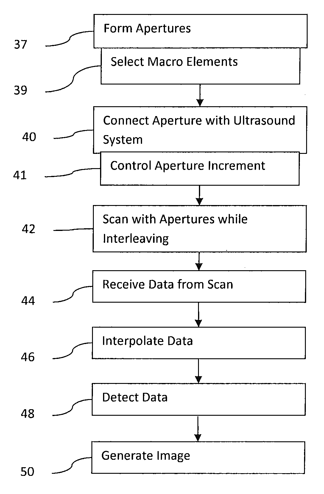
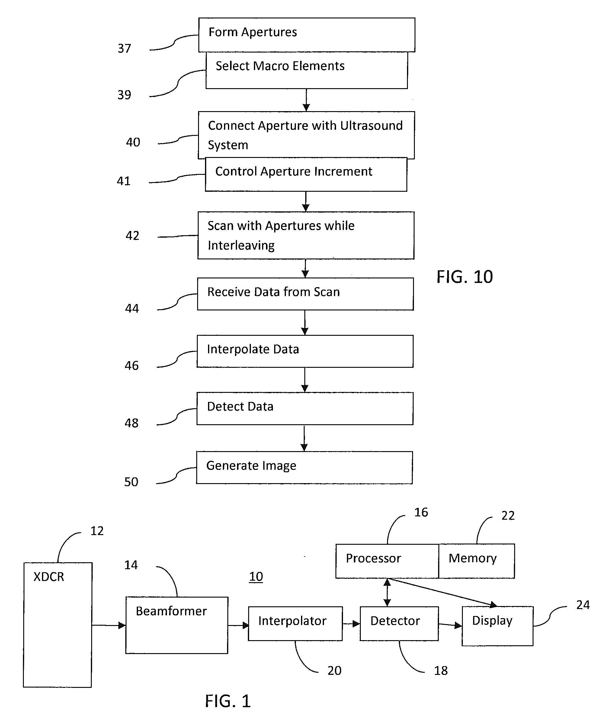
[0025]Electronically rotatable one-dimensional ultrasound arrays may provide a simplified and low cost real-time three-dimensional volume acquisition suitable for cardiac or other imaging. Fewer beamforming channels may be needed than scanning with a fully sampled two-dimensional array.

[0026]A randomized or aperiodic cell structure provides better beam quality than a periodic structure. An acoustic cell pitch less than or equal to half the macro element width may allow for aperiodic macro elements in addition to or alternative to aperiodic cell structure alone. In one embodiment, a coarse periodic electronic cell grid is combined with a finer aperiodic acoustic cell grid for increasing imaging performance.

[0027]In one embodiment, a reconfigurable array architecture has an electronic cell matrix interconnected laterally by a first set of switches and an overlaid acoustic cell matrix connected “vertically” to nearby electronic cells by a second set of switches. The electronic cell mat...


PUM


Abstract

Medical diagnostic ultrasound imaging is performed with a multi-dimensional transducer array and an imaging system for planar scanning. The elements of the array may be distributed on a periodic grid with aperiodic shifts in position. When a one-dimensional array is formed on the array, the aperiodic shifts better distribute acoustic energies, reducing peaks in side lobes. Using a layered structure of switches underneath the acoustic elements, side lobes may be further reduced. The switches are used for interconnecting elements to form macro elements of the one-dimensional aperture on the multi-dimensional array. The switches are distributed on a grid corresponding to the desired imaging frequency. The acoustic elements are distributed with a finer pitch. The finer pitch allows formation of the macro elements for the one-dimensional aperture where the edges of the macro elements have fewer or no periodic patterns.

Description

RELATED APPLICATIONS[0001]The present patent document claims the benefit of the filing date under 35 U.S.C. §119(e) of Provisional U.S. Patent Application Ser. No. 61 / 227,639, filed Jul. 22, 2009, which is hereby incorporated by reference.BACKGROUND[0002]The present embodiments relate to ultrasound transducers. In particular, the present embodiments relate to a reconfigurable transducer array.[0003]One-dimensional transducers are used to scan a plane with electronic steering. For scanning a volume, a two-dimensional transducer may be used. However, the number of beamforming channels greatly increases for scanning with a two-dimensional transducer. Many ultrasound imaging systems do not have sufficient channels. An alternative is a mechanically rotated one-dimensional transducer (i.e., a wobbler). However, the mechanical motion may be insufficiently rapid for real-time volume scanning.[0004]A multi-dimensional transducer array may be used with electronic switching to both provide for...

Claims


Application Information

Patent Timeline
Patent Type & Authority Applications(United States)
IPC IPC(8): A61B8/14
CPCA61B8/54G01N29/0654G01N29/245A61B8/483G01S7/52085G01S15/8925G01S15/8927G01N29/262
Inventor DAFT, CHRISTOPHER M.WAGNER, PAUL A.
Owner SIEMENS MEDICAL SOLUTIONS USA INC
Why Eureka
Try Eureka