Imaging and evaluating embryos, oocytes, and stem cells

a technology of embryos and stem cells, applied in the field of biological and clinical testing, can solve the problems of low birth rate, miscarriage, and well-documented adverse outcomes for both mother and fetus, and achieve good developmental potential, good developmental potential, and good developmental potential

Active Publication Date: 2011-04-21
THE BOARD OF TRUSTEES OF THE LELAND STANFORD JUNIOR UNIV
View PDF31 Cites 86 Cited by
  • Summary
  • Abstract
  • Description
  • Claims
  • Application Information

AI Technical Summary

Problems solved by technology

Infertility is a common health problem that affects 10-15% of couples of reproductive-age.
Multiple gestations have well-documented adverse outcomes for both the mother and fetuses, such as miscarriage, pre-term birth, and low birth rate.
Potential causes for failure of IVF are diverse; however, since the introduction of IVF in 1978, one of the major challenges has been to identify the embryos that are most suitable for transfer and most likely to result in term pregnancy.
The understanding in the art of basic embryo development is limited as studies on human embryo biology remain challenging and often exempt from research funding.
These differences, and many others make it inappropriate to directly extrapolate from one species to another.
(2009) Fertil. Steril.) In spite of such differences, the majority of studies of preimplantation embryo development derive from model organisms and are difficult to relate to human embryo development (Zernicka-Goetz, M.
However, potential risks of these methods also exist in that they prolong the culture period and disrupt embryo integrity (Manipalviratn S, et al.
However, these methods do not recognize the importance of the duration of cytokinesis or time intervals between early divisions.
However, these methods disclose only a basic and general method for time-lapse imaging of bovine embryos, which are substantially different from human embryos in terms of developmental potential, morphological behavior, molecular and epigenetic programs, and timing and parameters surrounding transfer.
Moreover, no specific imaging parameters or time intervals are disclosed that might be predictive of human embryo viability.
However, this work concluded that early first cleavage was not an important predictive parameter, which contradicts previous studies (Fenwick, et al.
Finally, no studies have validated the imaging parameters through correlation with the molecular programs or chromosomal composition of the embryos.

Method used


Image

  • Imaging and evaluating embryos, oocytes, and stem cells
  • Imaging and evaluating embryos, oocytes, and stem cells
  • Imaging and evaluating embryos, oocytes, and stem cells

Examples


example 1

[0167]Imaging analysis to determine developmental potential of embryos.

Methods

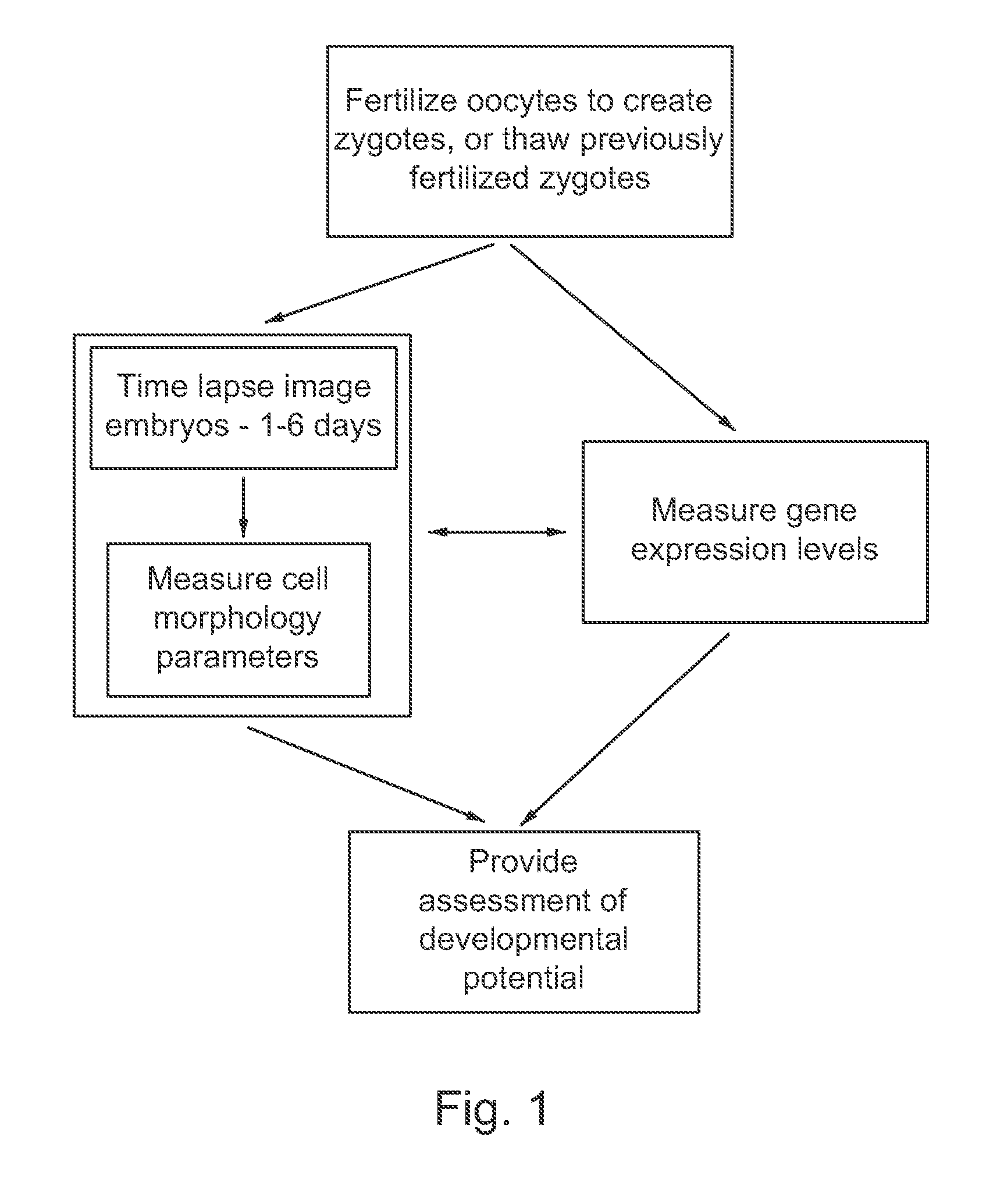
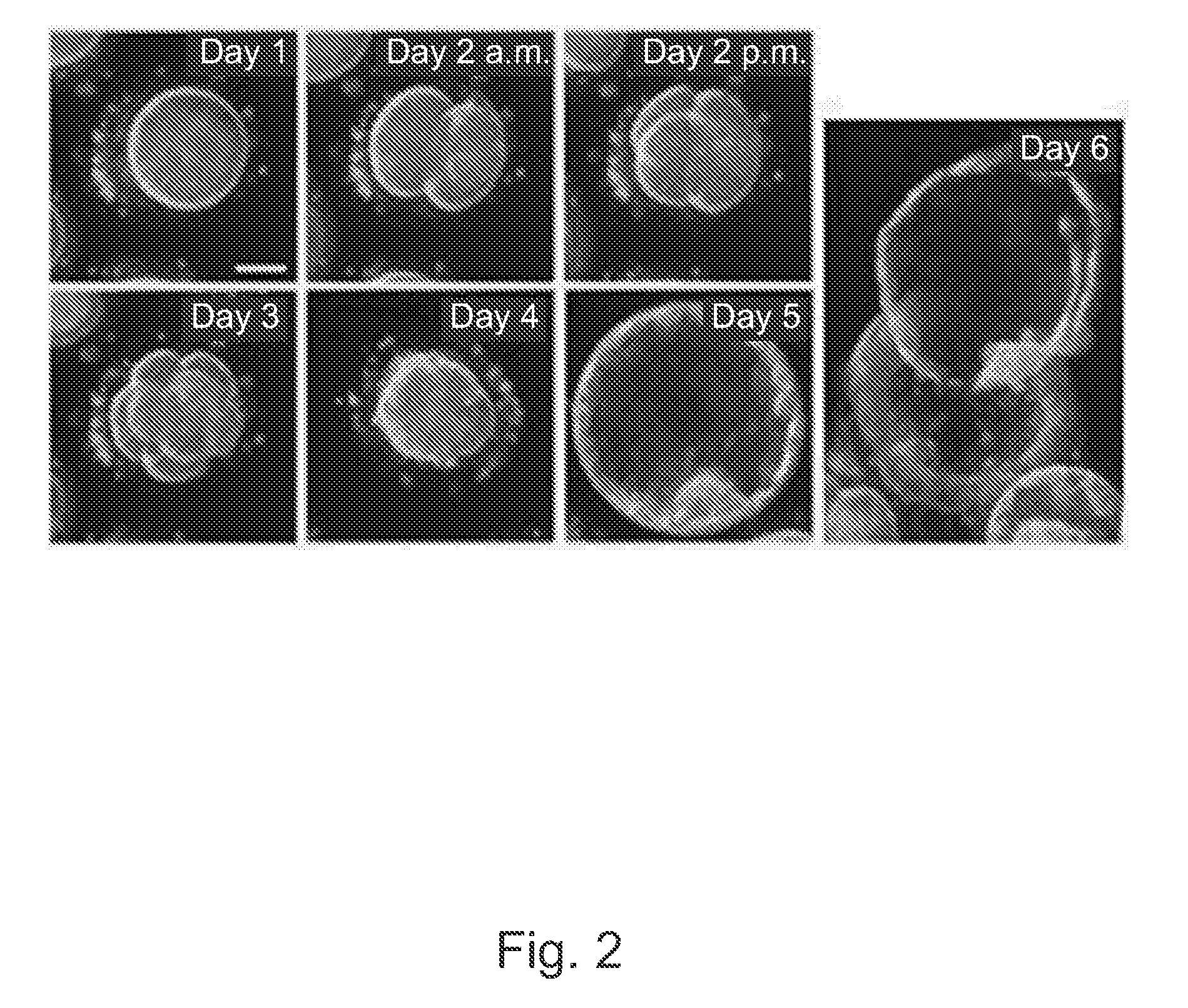
[0168]Frozen 1-cell human embryos, also referred to as zygotes, were thawed and placed into culture and cultured under conditions such as those used in IVF procedures. As described in more detail above, these embryos appear to be representative of the typical in vitro fertilization (IVF) population as they were frozen at the 2PN stage and thus indiscriminately cryopreserved. This is in contrast to embryos typically cryopreserved at later stages of development following transfer of those perceived to be of the highest quality during fresh cycles. For some experiments, embryos were placed in a standard culture dish. For other experiments, embryos were cultured in custom culture dish with optical quality micro-wells.

[0169]The growing embryos, typically between 1 to 30 per dish, were followed individually by time lapse imaging with a computer controlled microscope equipped for digital image storage and analysi...

example 2

[0178]Validation of imaging parameters through gene expression analysis, and use of gene expression analysis to determine developmental potential.

Methods

[0179]Frozen 1-cell human embryos, also referred to as zygotes, were thawed and placed into culture and cultured under conditions such as those used in IVF procedures. For some experiments, embryos were placed in a standard culture dish. For other experiments, embryos were cultured in custom culture dish with optical quality micro-wells.

[0180]Embryos were removed from the culture and imaging system and collected as either single embryos or single cells (blastomeres) for gene expression analysis. Each plate typically contained a mixture of embryos, with some reaching the expected developmental stage at the time of harvest, and others arresting at earlier developmental stages or fragmenting extensively. Those that reached the expected developmental stage at the time of harvest were classified as “normal”, whereas those that arrested w...

example 3

[0195]Imaging oocyte maturation and subsequent embryo development.

Results

[0196]One of the major limitations of current IVF procedures is oocyte quality and availability. For example, current IVF protocols recruit oocytes from the small cyclic pool, providing a small number of oocytes (e.g. 1-20) for fertilization. Moreover, approximately 20% of oocytes retrieved following hormone stimulation during IVF procedures are classified as immature, and are typically discarded due to a reduced potential for embryo development under current culture conditions.

[0197]One method to increase the oocyte pool is through in vitro maturation. FIG. 14 shows three stages of development during in vitro maturation, including germinal vesicle, metaphase I, and metaphase II. The germinal vesicle and metaphase I stages are classified as immature oocytes, while metaphase II is classified as mature due to the presence of the first polar body, which occurs at 24-48 hours after initiating in vitro maturation. F...


PUM

PropertyMeasurementUnit
timeaaaaaaaaaa
timeaaaaaaaaaa
time-aaaaaaaaaa

Abstract

Methods, compositions and kits for determining the developmental potential of one or more embryos or pluripotent cells and / or the presence of chromosomal abnormalities in one or more embryos or pluripotent cells are provided. These methods, compositions and kits find use in identifying embryos and oocytes in vitro that are most useful in treating infertility in humans.

Description

CROSS REFERENCE TO RELATED APPLICATIONS[0001]This application claims priority to U.S. Provisional Patent Application No. 61 / 332,651, filed May 7, 2010 and U.S. Provisional Patent Application No. 61 / 236,085, filed Aug. 22, 2009, both of which are herein incorporated by reference in their entireties.FIELD OF THE INVENTION[0002]This invention relates to the field of biological and clinical testing, and particularly the imaging and evaluation of zygotes / embryos, oocytes, and stem cells from both humans and animals.BACKGROUND OF THE INVENTION[0003]Infertility is a common health problem that affects 10-15% of couples of reproductive-age. In the United States alone in the year 2006, approximately 140,000 cycles of in vitro fertilization (IVF) were performed (cdc.gov / art). This resulted in the culture of more than a million embryos annually with variable, and often ill-defined, potential for implantation and development to term. The live birth rate, per cycle, following IVF was just 29%, wh...

Claims


Application Information

Patent Timeline
Patent Type & Authority Applications(United States)
IPC IPC(8): A61B17/435C12Q1/02C12Q1/68C12M1/34C12M1/22G16B45/00
CPCC12N5/0604G06K9/00147G02B21/0004C12Q1/02C12M41/48C12M21/06C12M41/36C12M41/46A61B17/435G06V20/698G16B45/00A61P15/08G01N15/1475G01N33/5005G01N2015/1497G01N2015/1493C12Q1/6881C12Q2600/158G01N15/10G01N2015/1087G01N2015/1093G06T7/0012G06T2207/10004G06T2207/10056G06T2207/30044
Inventor WONG, CONNIE C.LOEWKE, KEVIN E.BAER, THOMAS M.REIJO-PERA, RENEE A.BEHR, BARRY
Owner THE BOARD OF TRUSTEES OF THE LELAND STANFORD JUNIOR UNIV
Why Eureka
Try Eureka