
Image processing apparatus, image processing program, storage medium and ultra-sonograph

a technology of image processing and image processing program, which is applied in the field of image processing apparatus, image processing program, storage medium and ultrasonic diagnostic equipment, can solve the problems of difficult to cure arteriosclerosis, thin imt, difficult to measure, and difficult to find symptoms, so as to reduce the margin of error

Inactive Publication Date: 2011-05-12
GIFU UNIVERSITY
View PDF10 Cites 4 Cited by
  • Summary
  • Abstract
  • Description
  • Claims
  • Application Information

AI Technical Summary

Benefits of technology

[0018]To achieve the foregoing objectives, a first aspect of the present invention provides an image processing apparatus comprising: carotid artery wall estimation means that acquires ultrasound B-mode images of consecutive frames to estimate a carotid artery wall at a carotid artery minor axis cross section included in an ultrasound B-mode image of a predetermined frame; and heart rate variability tracking means that uses, as a template image, the ultrasound B-mode image including the estimated carotid artery wall and surrounding tissues of the carotid artery wall and estimates the size of the diameter of the carotid artery wall so as to minimize a margin of error between a transformed template, which is created by transforming the template image, and the acquired ultrasound B-mode image of each frame, thereby acquiring a time change of the carotid artery wall.
[0019]Preferably, the carotid artery wall estimation means determines the value of a carotid artery parameter for determining the position and the shape of the carotid artery so as to maximize an evaluation function indicating a degree of probability of a carotid artery, thereby estimating the carotid artery wall.
[0021]A second aspect of the present invention provides an image processing program causing a computer to function as: carotid artery wall estimation means that acquires ultrasound B-mode images of consecutive frames to estimate a carotid artery wall at a carotid artery minor axis cross section included in an ultrasound B-mode image of a predetermined frame; and heart rate variability tracking means that uses, as a template image, the ultrasound B-mode image including the estimated carotid artery wall and surrounding tissues of the carotid artery wall and estimates the size of the diameter of the carotid artery wall so as to minimize a margin of error between a transformed template image, which is created by transforming the template image, and the acquired ultrasound B-mode image of each frame, thereby acquiring a time change of the carotid artery wall.
[0022]Preferably, the carotid artery wall estimation means determines the value of a carotid artery parameter for determining the position and the shape of the carotid artery so as to maximize an evaluation function indicating a degree of probability of a carotid artery, thereby estimating the carotid artery wall.

Problems solved by technology

It is difficult to cure arteriosclerosis.
However, at an early stage of arteriosclerosis, there is no plaque, and the IMT is thin and hard to measure.
It is difficult to find symptoms at an early stage in such a morphological observation.
Although satisfied in many cases, the precondition is not satisfied in a case where there is local arteriosclerosis in the carotid artery.
Therefore, in such a case, the method previously proposed by the present applicant cannot be applied.
However, when a minor axis cross-sectional image of a carotid artery is used, it is difficult to use information related to a spatial shape of the image to improve the estimation accuracy due to the following reasons.
Therefore, it is difficult to express the shapes uniformly.
Therefore, usable information is scarce.
However, such a method for improving the estimation accuracy cannot be applied to a method of using a B-mode image to estimate and track the movement of a carotid artery.
However, ultrasonic diagnostic equipment capable of outputting RF signals is limited to only some models.

Method used


Image

  • Image processing apparatus, image processing program, storage medium and ultra-sonograph
  • Image processing apparatus, image processing program, storage medium and ultra-sonograph
  • Image processing apparatus, image processing program, storage medium and ultra-sonograph

Examples


embodiment

Operation of Embodiment

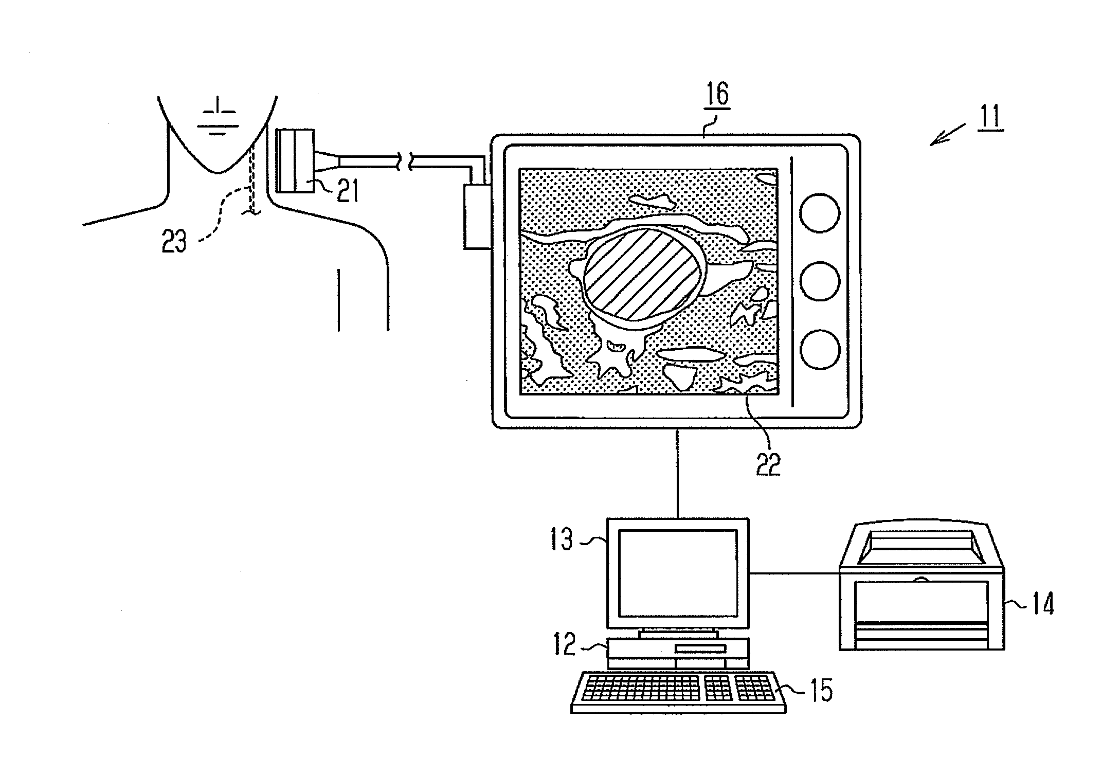
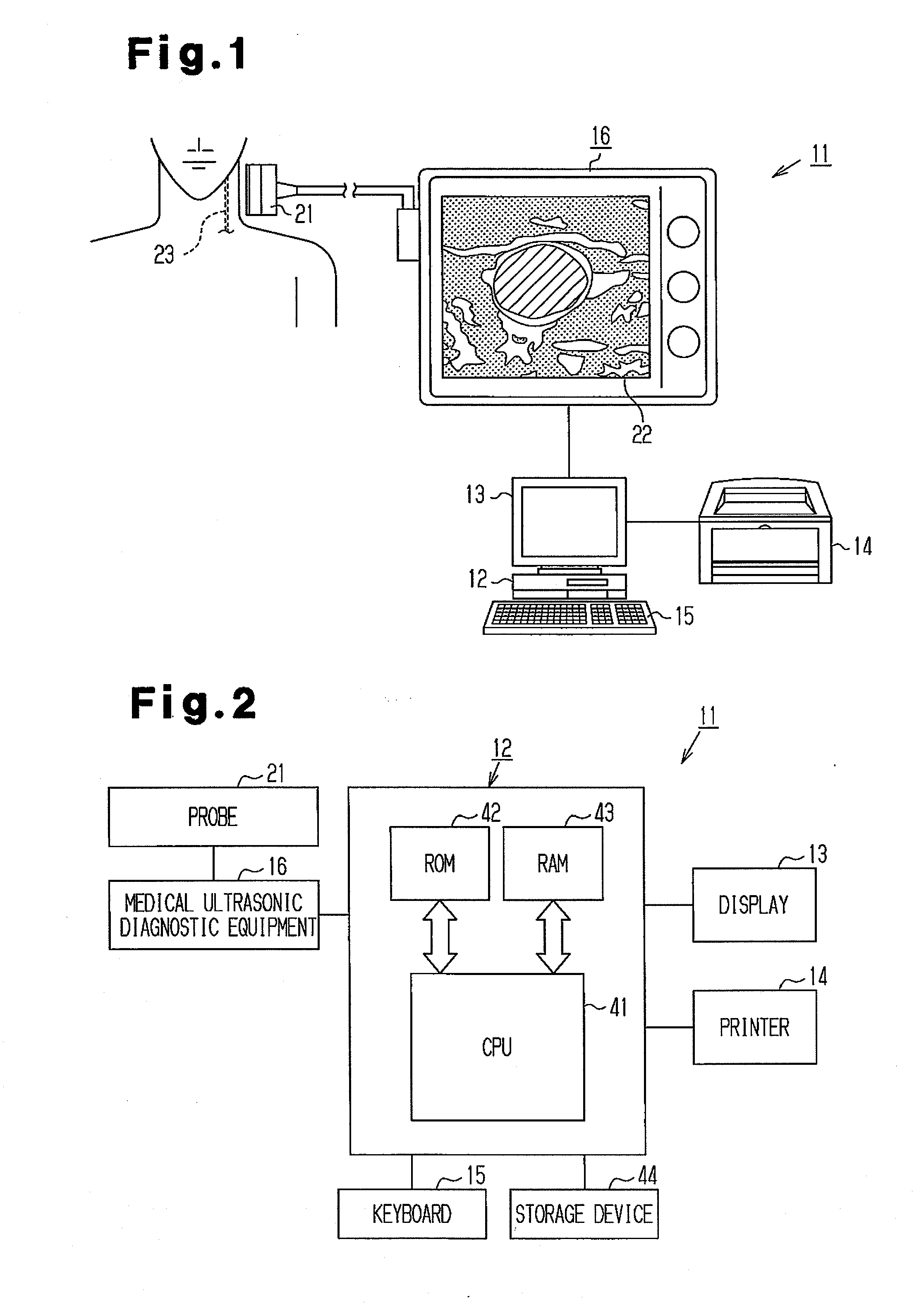
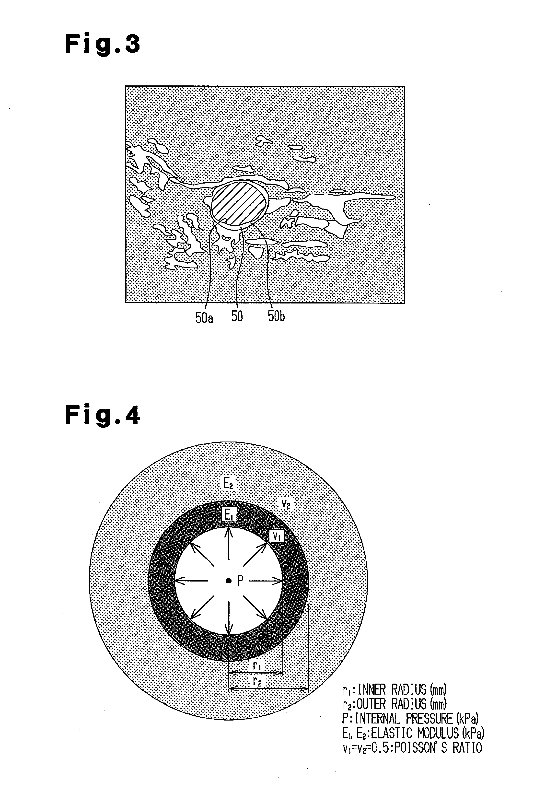
[0046]Next, a process of the image processing program executed by the CPU 41 in the image processing apparatus 11 configured as described above will be described. Before the description of the program processing, “stress and distortion characteristics of carotid artery and surrounding tissues”, “two-layer cylindrical model”, and “summary of method for tracking heart rate variability of carotid artery wall” will be described.

(Stress and Distortion Characteristics of Carotid Artery and Surrounding Tissues)

[0047]Carotid arteries repeat expansion and contraction by heartbeats. Surrounding tissues, such as fat, around the carotid arteries expand and contract in association with the expansion and contraction of the carotid arteries. In general, the relationship between the stress and distortion of substance is examined from the perspective of strength of materials.

[0048]FIG. 3 shows an ultrasound B-mode image (hereinafter, also simply referred to as “image”) includi...


PUM


Abstract

A computer 12 of an image processing apparatus 11 acquires ultrasound B-mode images of consecutive frames to estimate a carotid artery wall at a carotid artery minor axis cross section included in an ultrasound B-mode image of a predetermined frame. The computer 12 also uses, as a template image, the ultrasound B-mode image, which includes the estimated carotid artery wall and surrounding tissues of the carotid artery wall and estimates the size of the diameter of the carotid artery wall so as to minimize a margin of error between a transformed template image, which is created by transforming the template image, and the acquired ultrasound B-mode image of each frame, thereby acquiring the time change of the carotid artery wall.

Description

TECHNICAL FIELD[0001]The present invention relates to an image processing apparatus, an image processing program, a storage medium, and ultrasonic diagnostic equipment, and particularly, to an image processing apparatus and an image processing program for an ultrasound image of a carotid artery as well as a storage medium and ultrasonic diagnostic equipment related to the image processing apparatus and the image processing program.BACKGROUND ART[0002]Arteriosclerosis is one of the lifestyle-related diseases in which excessive lifestyle habits are risk factors. Arteriosclerosis advances from youth with very few symptoms, and if left untreated, arteriosclerosis may develop into cerebral infarction, cardiac infarction, angina pectoris, and the like. It is difficult to cure arteriosclerosis. Therefore, it is important to find symptoms of arteriosclerosis at an early stage and prompt the patients to improve lifestyle habits to slow down the progression of the diseases.[0003]There are var...

Claims


Application Information

Patent Timeline
IPC IPC(8): A61B8/14G06K9/00
CPCA61B5/02007A61B8/0858G01S7/52042A61B8/485A61B8/463
Inventor YOKOTA, YASUNARINOGATA, FUMIOKAWAMURA, YOKO
Owner GIFU UNIVERSITY
Learn More