Phage Display Using Cotranslational Translocation of Fusion Polypeptides

a polypeptide and cotranslational translocation technology, applied in the field of phage display method, can solve the problems of polypeptide recalcitrant display, inability to fully explore the cotranslational translocation pathway, and inability to fully explain the contribution of the translocation mechanism to the success of phage display

Active Publication Date: 2011-05-19
UNIV ZURICH
View PDF0 Cites 39 Cited by
  • Summary
  • Abstract
  • Description
  • Claims
  • Application Information

AI Technical Summary

Benefits of technology

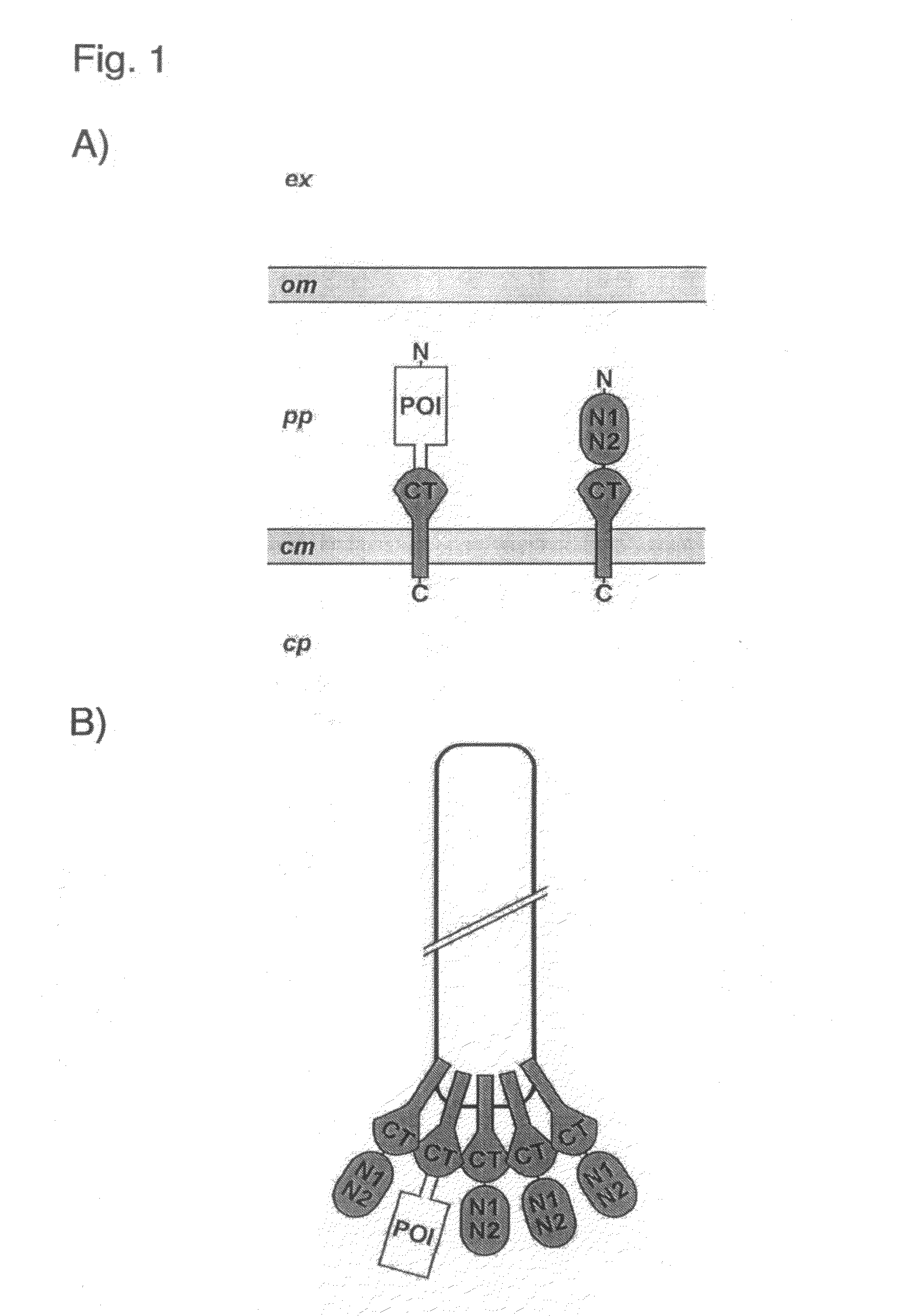
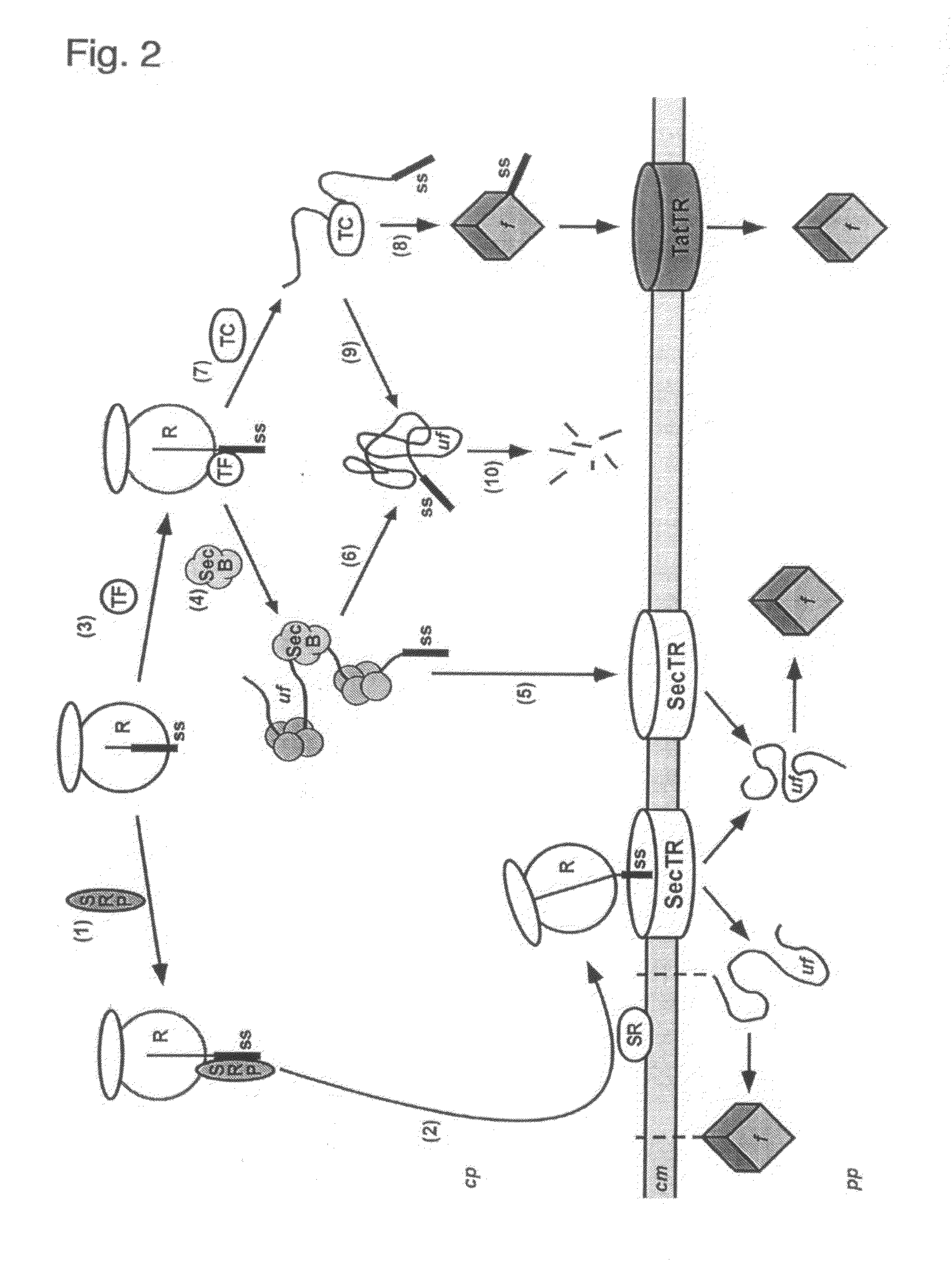
[0010]Accordingly, the present invention allows phage display by cotranslational translocation of the fusion polypeptides comprising the POI. This method is particularly suitable for polypeptides, w

Problems solved by technology

However, the contribution of the mechanism of translocation for the success of phage display has not been fully elucidated, and the possibility to use a cotranslational translocation pathway was not explored in the prior art.
Nevertheless, some polypeptides are recalcitrant to display due to individual properties, mostly because of unknown reasons.
This makes the success of the display of a certain protein unpredictable.
In addition, when a combinatorial library is created for phage display, not all clones will display with similar efficiency; this is

Method used


Image

  • Phage Display Using Cotranslational Translocation of Fusion Polypeptides
  • Phage Display Using Cotranslational Translocation of Fusion Polypeptides
  • Phage Display Using Cotranslational Translocation of Fusion Polypeptides

Examples


example

[0086]Materials

[0087]Chemicals were purchased from Fluka (Switzerland). Oligonucleotides were from Microsynth (Switzerland). Vent DNA polymerase, restriction enzymes and buffers were from New England Biolabs (USA) or Fermentas (Lithuania). Helper phage VCS M13 was from Stratagene (USA). All cloning and phage amplification was performed in E. coli XL1-Blue from Stratagene (USA).

[0088]Molecular Biology

[0089]Unless stated otherwise, all molecular biology methods were performed according to described protocols (Ausubel, F. M., Brent, R., Kingston, R. E., Moore, D. D., Sedman, J. G., Smith, J. A. and Stuhl, K. eds., Current Protocols in Molecular Biology, New York: John Wiley and Sons, 1999). Brief protocols are given below.

[0090]Phage Display Related Methods

[0091]Unless stated otherwise, all phage display related methods were performed according to described protocols (Clackson, T. and. Lowman, H. B. eds., Phage Display A Practical Approach, New York: Oxford University Press, 2004; Barb...


PUM


Abstract

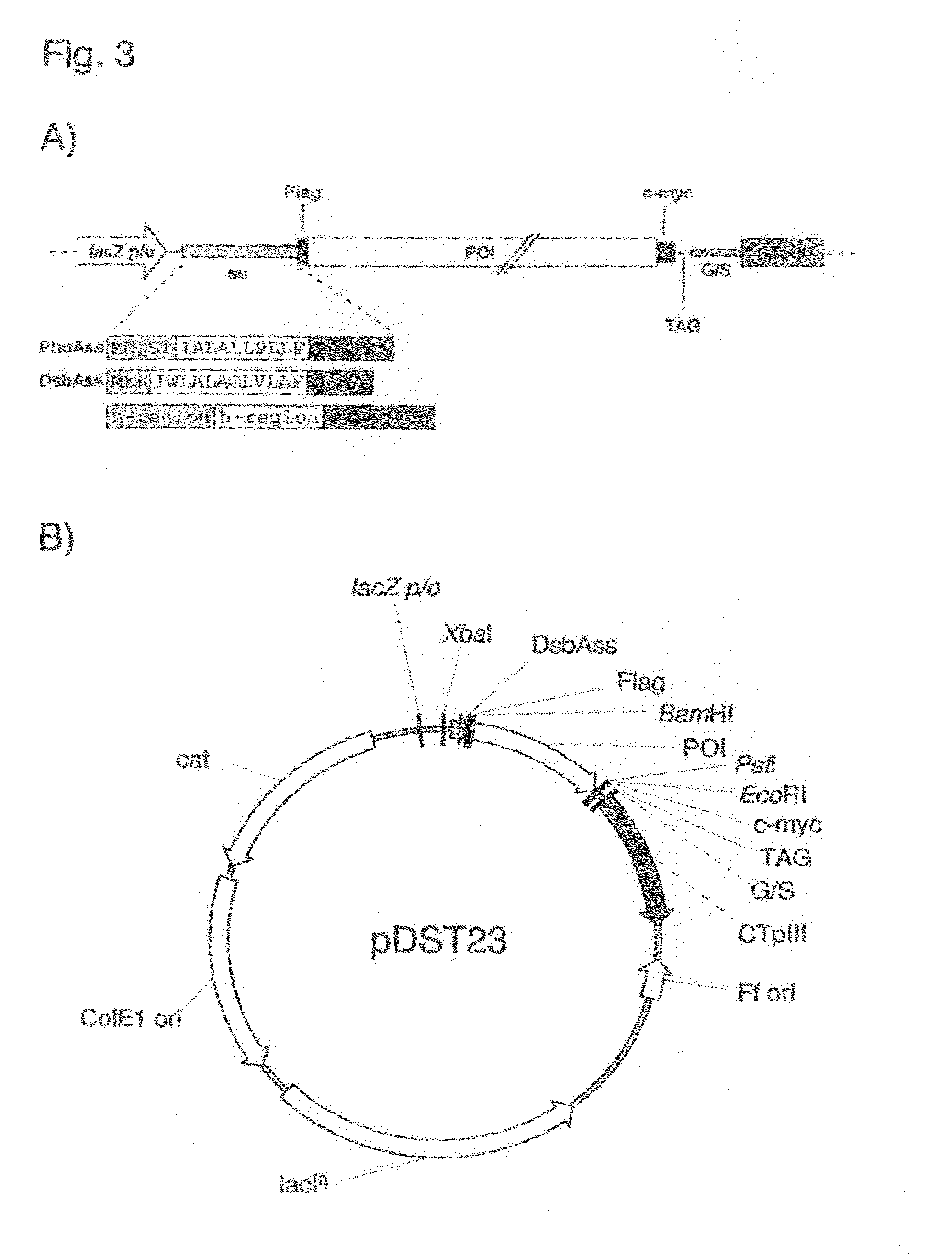
The present invention relates to a filamentous phage display method wherein the polypeptides of interest displayed on the phage particle are cotranslationally translocated across the cytoplasmic membrane of Gram-negative bacteria based on the signal recognition particle pathway. This method is particularly suitable for polypeptides, which are known to be difficult to display on phages, and for proteins of cDNA libraries and other combinatorial libraries, in particular when derived from very fast folding, stable protein scaffolds. The invention further relates to phage or phagemid vectors useful in the method comprising a gene construct coding for a fusion polypeptide comprising the polypeptide to be displayed on the phage particle and an N-terminal signal sequence promoting cotranslational translocation.

Description

FIELD OF THE INVENTION [0001]The present invention relates to a novel phage display method, phage or phagemid vectors used therein and the phage particles so obtained.BACKGROUND OF THE INVENTION[0002]Display of polypeptides on bacteriophage (phage display) is a selection technique that allows to extract polypeptides with desired properties from a large collection of variants (Russel, M., Lowman, H. B., and Clackson, T., Introduction to phage biology and phage display, in “Phage Display”; Clackson, T. and Lowman, H. B., eds., Oxford University Press, 2004, pp. 1-26). Phage display has been intensively investigated for the selection from combinatorial antibody or peptide libraries.[0003]By far the most widely used bacteriophages used in phage display are filamentous phages. Filamentous phages constitute a large family of bacterial viruses that infect many Gram-negative bacteria. The best-known filamentous phages are those that infect Escherichia coli; these are f1 / M13 / fd and IKe. Phag...

Claims


Application Information

Patent Timeline
IPC IPC(8): C40B40/02C40B50/06C12N15/63
CPCC07K2319/034C07K2319/43C07K2319/50C12N15/63C12N15/1037C07K1/047C07K2319/735C12N15/625C40B40/02C12N15/10C12N7/00
Inventor STEINER, DANIELFORRER, PATRIKSTUMPP, MICHAEL T.PLUCKTHUN, ANDREAS
Owner UNIV ZURICH
Why Eureka
Try Eureka