Assay system to identify therapeutic agents

a technology of assay system and therapeutic agent, applied in the field of assay system, to achieve the effect of facilitating the identification or qualification of pore formation, and reducing the binding of liposome reagen

Active Publication Date: 2011-06-02
EUTROPICS PHARMA
View PDF7 Cites 39 Cited by
  • Summary
  • Abstract
  • Description
  • Claims
  • Application Information

AI Technical Summary

Benefits of technology

[0009]A liposome reagent contains a liposome in which a BCL-2 family protein attached to said liposome and a detection reagent encapsulated within the liposome. The liposome is composed of vesicle forming lipids such as phosphatidyl choline, phosphatidyl serine, phosphatidyl inositol or cardiolipin. Optionally, the liposome contains at least 10 mole percent of cardiolipin. Additionally, the liposome contains at least 1 mole percent of vesicle forming lipid that is derivatized with 4,4-difluoro-3a,4a-diaza-s-indacene (BODIPY). The liposome contain 2, 3, 4, 5 or more mole percent of BODIPY.
[0010]A detection reagent is any reagent suitable to facilitate the identification or qualification of pore formation in the liposome. For example, the detection reagent is a fluorochrome, a chromogen or a dye. Exemplary detection reagents include fluorescein dextran or coumeran dextran.
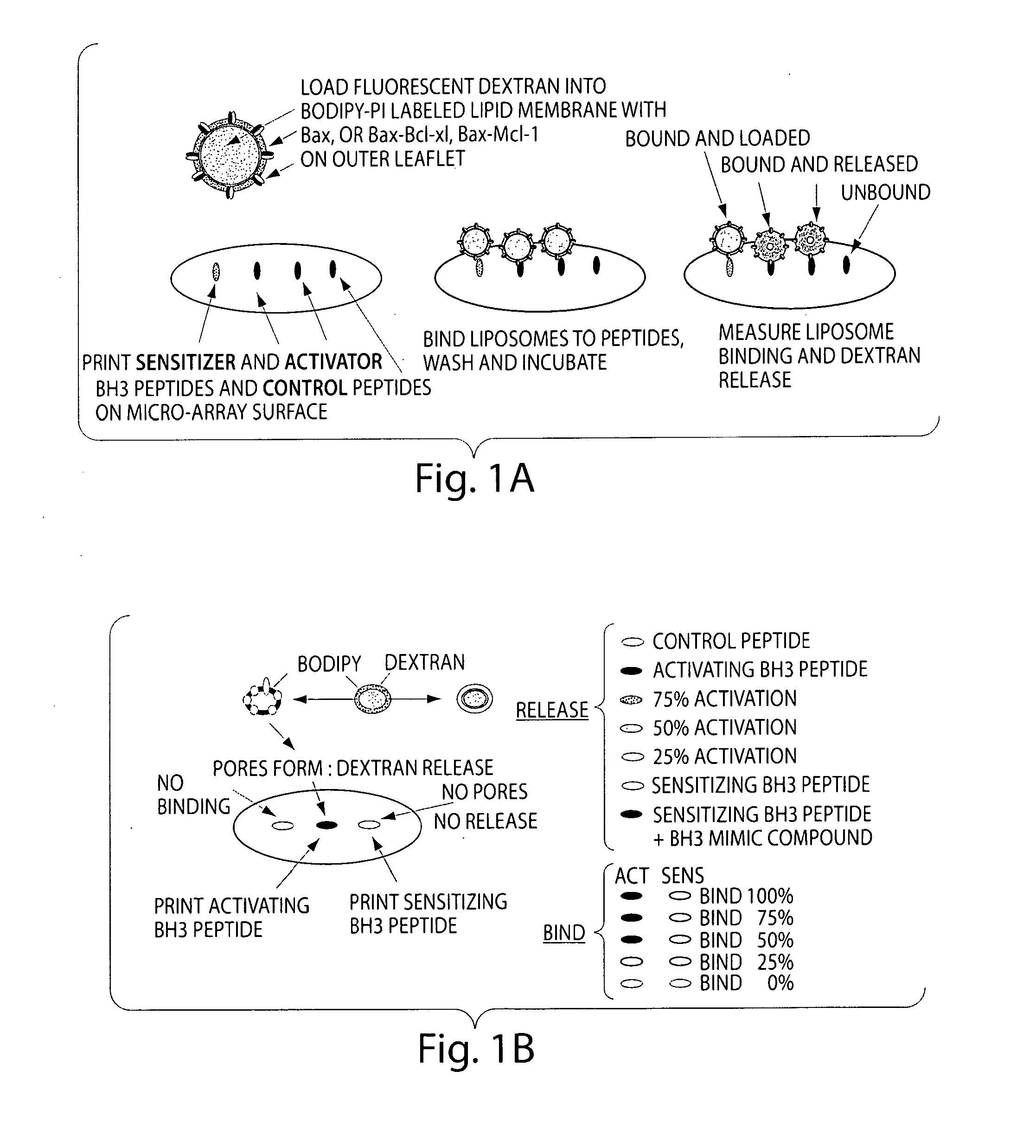
[0011]Also included in the invention are methods of method of identifying an activating BH3 and sensitizing mimetic by contacting a test compound with a liposome assay system according to the invention and detecting binding of the liposome reagent to the solid substrate and release of the detection reagent from the liposome. A decrease in binding of the liposome reagent in the presence of the test compound compared to the absence of the test compound and release of the detection reagent indicates that the test compound is an activating BH3 mimetic. In contrast, a decrease in binding of the liposome reagent in the presence of the test compound compared to the absence of the test compound and no release of the detection reagent from the liposome indicates that the test compound is a sensitizing BH3 mimetic. Also provided by the invention are methods of predicting sensitivity of a cancer cell or subject to a therapeutic agent by contacting a mitochondria that has been isolated from a cancer cell from a subject and labeled with a potentiometric dye and a solid substrate containing and immobilized BH3 domain peptide and a mitochondrial binding agent under conditions where the mitochondria and mitochondrial binding agent are capable of forming a complex. The complex is contacted with a test therapeutic agent and the release of the potentiometric dye from the mitochondria is detected. Optionally, the complex is washed prior to contacting the complex with the test therapeutic agent or the complex is labeled prior to detecting potentiometric dye release. An increase in release of the potentiometric dye from the mitochondria in the presence of the test therapeutic agent compared to the absence of the test therapeutic agent indicates that the cancer cell or subject is sensitive to the therapeutic agent.
[0012]Unless otherwise defined, all technical and scientific terms used herein have the same meaning as commonly understood by one of ordinary skill in the art to which this invention belongs. Although methods and materials similar or equivalent to those described herein can be used in the practice or testing of the present invention, suitable methods and materials are described below. All publications, patent applications, patents, and other references mentioned herein are incorporated by reference in their entirety. In the case of conflict, the present specification, including definitions, will control. In addition, the materials, methods, and examples are illustrative only and are not intended to be limiting.

Problems solved by technology

A key challenge for the drug industry is to discover small molecules or peptides that block domain-mediated protein-protein interactions, and do so in a subset specific manor.

Method used


Image

  • Assay system to identify therapeutic agents
  • Assay system to identify therapeutic agents
  • Assay system to identify therapeutic agents

Examples


example 1

Two Color Vesicle Release Assay

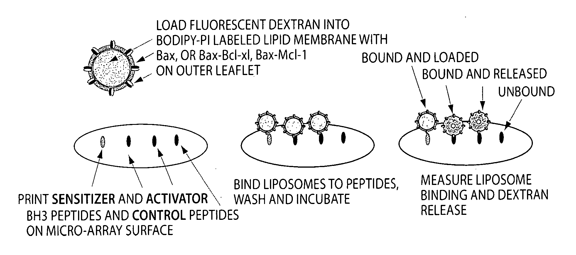
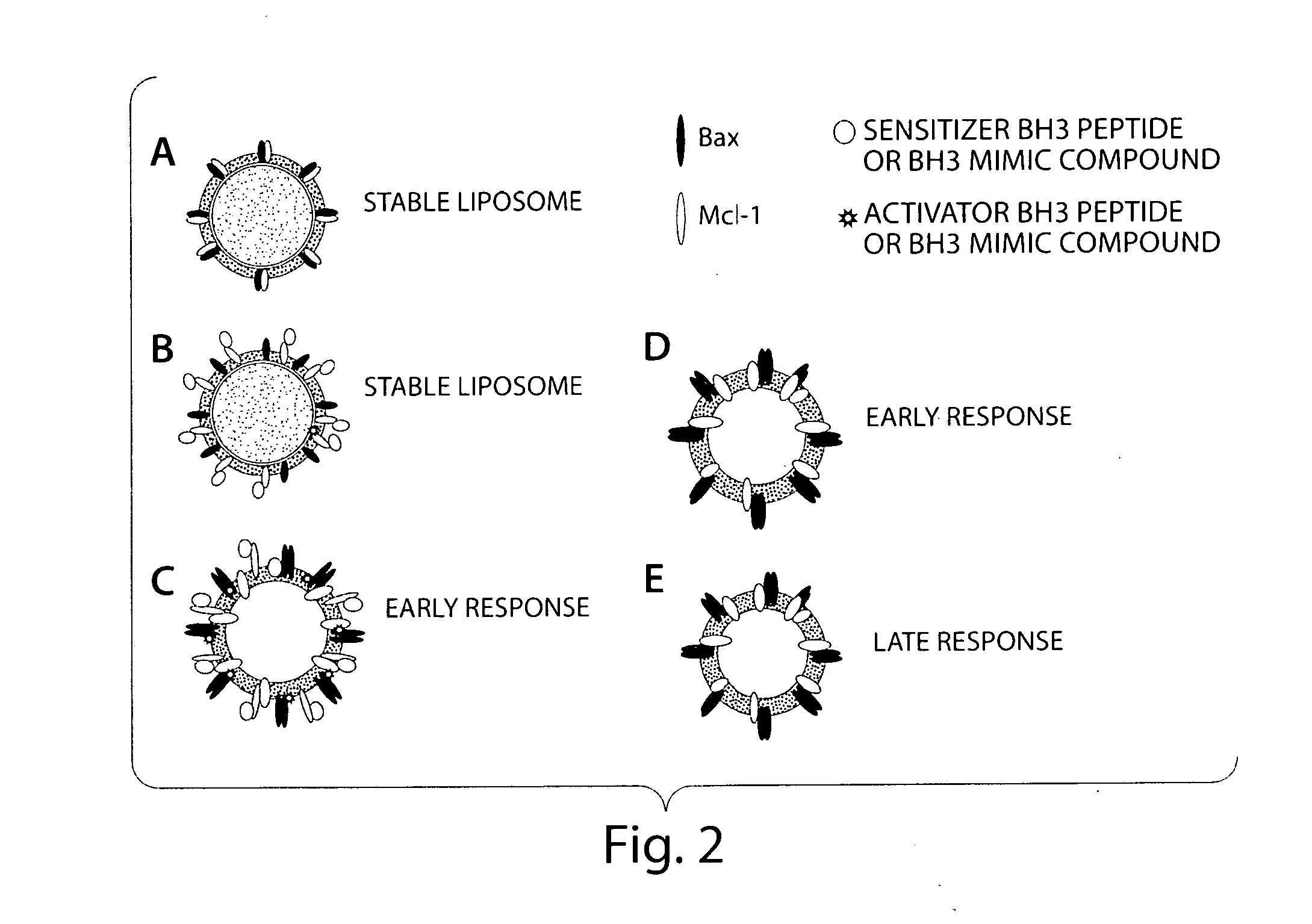
[0070]A simplified cell free system is used to detect Bax pore forming function on a lipid bilayer. The liposome release assay measures both the release of the fluorescently labeled contents and the lipid itself so a ratio of signals from each label or a shift in FRET will give accurate measurement of pore forming activity. Both a ratio of signals from each label and a shift in FRET are tested.

[0071]To accomplish this assay, purified proteins are incubated with synthetic liposomes labeled with BODIPY (either Phosphotidyl Inositol or Di-oc) and containing entrapped fluorescien dextran or coumeran dextran. The enhancement of Bax pore forming activity by BH3 peptide or BH3 mimic compounds is assessed in a liquid phase assay. Both colors will be read as well as the FRET signal generated from energy transfer between the fluorescence in the lumen of the liposome (coumeran or fluorescence) and that on the inner leaflet of the liposome. Efficiency is determine...

example 2

Assessment of Binding of the Two Color Lipid Vesicles to Printed BH3 Peptide Array

[0079]A peptide array format is used to assess the binding of Bax vesicles. These arrays are highly sensitive at detecting non-lipid associated Bcl-xL, Bcl-2 and Bax binding. Assays are prepared as described below, except that the format is changed to accommodate the traditional microscope slide. Using the microscope slide for a rough determination of a read out is sufficient for a first level experiment to identify that the system works. In a second level of experimentation, the format described above in Example 1 is used. Liposomes are prepared as described in Example 1, and incubated on the arrays and allowed to bind and incubated over a time course. Condition for optimal binding is determined by the liposome release profile and the readouts that indicate binding of molecules to the BH3 binding domains. Binding constants are calculated.

[0080]To generate the peptide arrays, peptides comprised of the ...

example 3

Optimization of Liposome Binding to Printed BH3 Peptides

[0081]Liposomes are labeled with combination of label determined in Example 1 above, and are prepared as described above. Liposome binding buffer includes PBS with other liposome compatible buffers. A phase 2 buffer is used without detergent, and the buffer specified in Kuwana et al can be used.

[0082]The Kds are established with purified proteins as a starting point and it is estimated that the amount of protein incorporated into a liposome is a direct function of the nature of the protein being used, and generally will agree with the amounts of protein incorporated into liposome for pore forming assays known in the art. The molar concentration of protein likewise follows generally that used for pore forming assays as described previously in the art.

[0083]Enough lipid is prepared to provide sufficient liposomes for the BH3 domain binding array, and is therefore loosely dependent on the amount of candidate therapeutic agents bei...


PUM

PropertyMeasurementUnit
diameteraaaaaaaaaa
diameteraaaaaaaaaa
number average sizesaaaaaaaaaa

Abstract

The invention is an assay designed to identify agents that modulate apoptosis. The invention provides an assay system and methods for screening for inhibitors of the Bcl-2 family of proteins. In various aspects the invention provides an assay system containing a liposome reagent and an immobilized BH3 domain peptide. In further aspects the invention provides an assay system containing mitochondria, an immobilized BH3 domain peptide and a mitochondrial binding agent, e.g. an anti-VDAC anti-body.

Description

RELATED APPLICATIONS[0001]This application claims the benefit of U.S. Ser. No. 60 / 838,284 filed Aug. 16, 2006 the contents of which is incorporated herein by reference in its entirety.FIELD OF THE INVENTION[0002]This invention relates generally to an assay system and methods for identifying modulators of apoptosis.BACKGROUND OF THE INVENTION[0003]The Bcl-2 (B cell lymphoma) family of proteins are genetically and functionally well characterized as being critical in effecting cellular apoptosis. Within this family are proteins that function either to protect cells against apoptotic signals or that induce apoptosis. A balance of interactions between the pro- and anti-apoptotic Bcl-2 family proteins determines whether the cell undergoes apoptosis or remains healthy. Bcl-2 is found to be over-expressed in many tumors including B-cell lymphomas, adenocarcinomas of the colon and prostate, gastric and naso-pharyngeal cancers. Bcl-2 has also been implicated in resistance of tumor cells to ch...

Claims


Application Information

Patent Timeline
Patent Type & Authority Applications(United States)
IPC IPC(8): C40B40/10G01N33/92
CPCA61K49/0004G01N2510/00G01N33/586G01N33/5079C40B40/10G01N33/53G01N33/543G01N33/574
Inventor CARDONE, MICHAEL H.
Owner EUTROPICS PHARMA
Why Eureka
Try Eureka