Flexible 3D microprobe structure

Active Publication Date: 2011-06-16
NATIONAL TSING HUA UNIVERSITY
View PDF3 Cites 13 Cited by
  • Summary
  • Abstract
  • Description
  • Claims
  • Application Information

AI Technical Summary

Benefits of technology

[0007]The primary objective of the present invention is to provide a flexible 3D (3-dimensional) microprobe structure, which adopts a flexible material having a better biocompatibility and less likely to cause inflammation, whereby the micropro

Problems solved by technology

A conventional planar MEA cannot fully record the electrophysiological signals.
Further, the neural signals from neurons need complicated time-consuming signal processing and computation.
If a hard material inserted into the tissues for a long time, the mechanical mismatch between the hard material and the soft tissues may cause inf

Method used


Image

  • Flexible 3D microprobe structure
  • Flexible 3D microprobe structure
  • Flexible 3D microprobe structure

Examples


Embodiment Construction

[0018]Below, the technical contents of the present invention are described in detail in cooperation with the drawings.

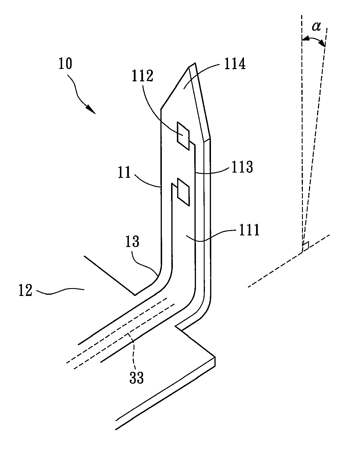
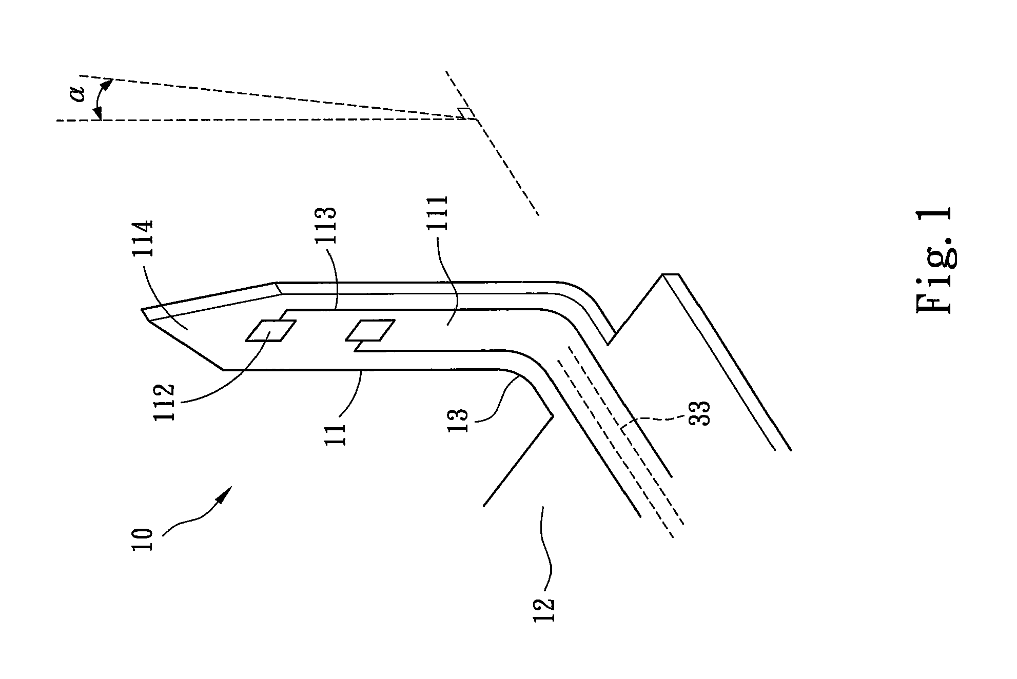
[0019]Refer to FIG. 1 a perspective view schematically showing the appearance of a flexible 3D microprobe structure 10 according to one embodiment of the present invention. The flexible 3D microprobe structure 10 comprises at least one probe 11, a base 12 and a hinge portion 13. The probe 11 is connected to the base 12 via the hinge portion 13. The probe 11 forms a bend angle α with respect to a normal of the base 12 by attracting the probe 11 through an electrostatic force to make the hinge portion 13 bend with respect to the base 12. The probe 11 and the hinge portion 13 are made of a first flexible polymeric material, and the base 12 is made of a second flexible polymeric material. The first and second materials are selected from a group consisting of polyimide (PI), parylene(poly-para-xylylen), a thick SU-8 photoresist, polydimethylsiloxane (PDMS), and benzocyclo...


PUM


Abstract

The present invention discloses a flexible 3D microprobe structure, which comprises at least one probe, a base and a hinge portion. The probe is connected to the base via the hinge portion. The probe forms a bend angle with respect to a normal of the base by attracting the probe through an electrostatic force to make the hinge portion bend with respect to the base, and thus to form a 3D structure having the bend angle. The probe, the base and the hinge portion are made of a flexible polymeric material to reduce the inflammation response of creatures. Further, a fixing element is used to enhance the structural strength of the flexible 3D microprobe structure.

Description

FIELD OF THE INVENTION[0001]The present invention relates to a flexible 3D microprobe structure, particularly to a flexible 3D microprobe structure formed by electrostatic force.BACKGROUND OF THE INVENTION[0002]Recently, advance in biomedical microelectronics has greatly improved in-vivo physiological research, for example, the studies of principles and methods of the neural conduction, which may favor the neurological diagnosis and treatment. The microelectrode array, or MEA for short, not only has fine puncturing ability but also can record in-vivo electrophysiological signals. Therefore, MEA plays a very important role in the field of neural science. Further, MEA also functions as an intermediary between analog physiological signal and bio signal analysis.[0003]A conventional planar MEA cannot fully record the electrophysiological signals. Thus, a 3D MEA is used to detect neural signals. In detection, MEA punctures tissue before detecting actional potentials of neuron, which may ...

Claims


Application Information

Patent Timeline
IPC IPC(8): A61B5/04
CPCA61B5/685A61B5/04001A61B5/24
Inventor YAO, DA-JENGCHEN, CHANG-HSIAOCHUANG, SHIH-CHANGCHANG, YEN-CHUNG
Owner NATIONAL TSING HUA UNIVERSITY
Why Eureka
Try Eureka